
|
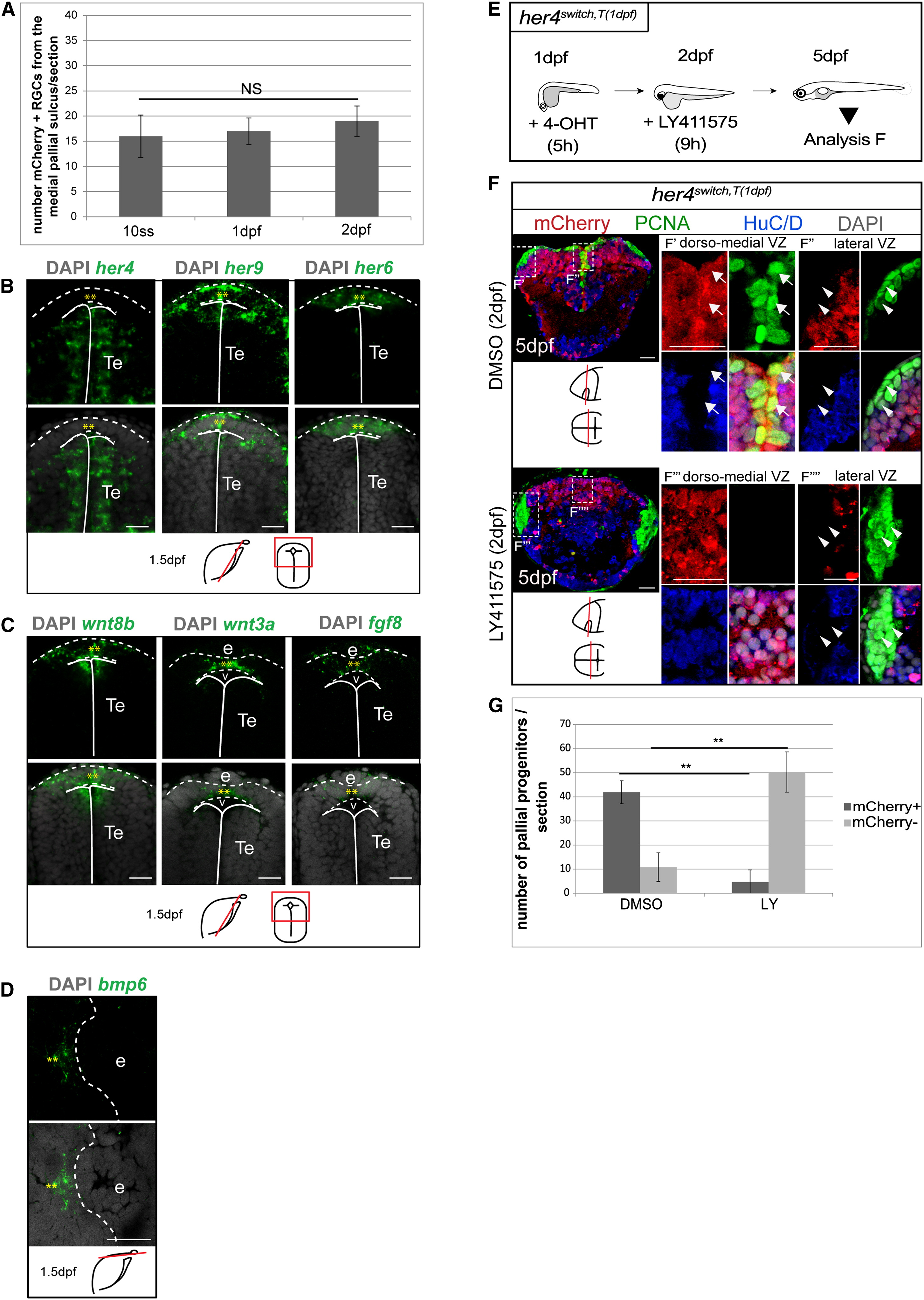
Fig. 4
The her4-Negative Cells at the Origin of Lateral Pallial aNSCs Belong to the “Progenitor Pool” Subclass of Embryonic Neural Progenitors
(A) Number of mCherry+ RGCs per adult telencephalic section, from the sulcus ypsiloniformis up to the edge of the VZ, after recombination at 1–10 somites, 1 dpf or 2 dpf of her4switch embryos. Values are presented as mean ± 95% CI (ANOVA).
(B–D) Compared expression of her4, her9, her6, wnt8b, wnt3a, fgf8, and bmp6 along the posterior telencephalic roof plate at 1.5 dpf, revealed with fluorescence in situ hybridization without/with 42,6-diamidino-2-phenylindole. Frontal (B and C) or horizontal (D) single confocal planes are shown. Dashed line represents roof plate of the neural tube (or epiphysis) and plain lines represent the ventricle. Roof plate. e, epiphysis; Te, telencephalon.
(E) Experimental design to assess Notch sensitivity of pallial progenitors at 2 dpf.
(F) Medial cross-sections of the telencephalon in her4switchT(1 dpf) larvae treated with DMSO or LY411575. Magnification of the dorsomedial VZ (F′ and F′′′) and lateral VZ (F′′ and F′′′′), immunostained as indicated. Arrows and arrowheads highlight, respectively, the dorsomedial progenitors (mCherry+/PCNA+ cells) and the lateral progenitors (mCherry/PCNA+ cells).
(G) Compared number of pallial dorsomedial progenitors (dark gray) and of lateral progenitors (light gray) in control (DMSO) and treated (LY411575) conditions. Values are presented as mean ± 95%CI (ANOVA, p<0.05).
See also Figure S4.
Reprinted from Developmental Cell, 30(2), Dirian, L., Galant, S., Coolen, M., Chen, W., Bedu, S., Houart, C., Bally-Cuif, L., Foucher, I., Spatial Regionalization and Heterochrony in the Formation of Adult Pallial Neural Stem Cells, 123-36, Copyright (2014) with permission from Elsevier. Full text @ Dev. Cell