
|
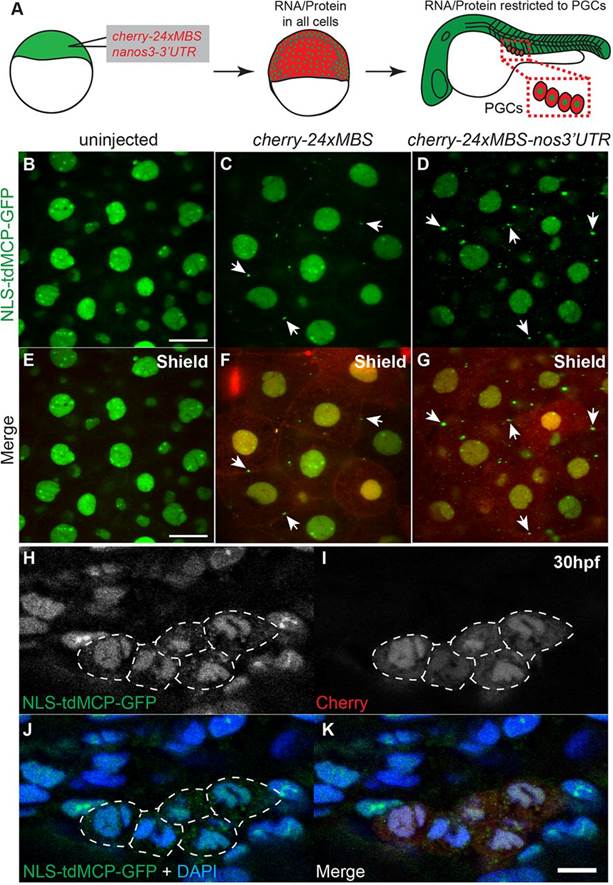
Fig. 3
MS2-tagged nanos3 3′UTR is detected by MCP-GFP in the cytoplasm of PGCs. (A) Schematic depicting injection and imaging of cherry-24xMBS-nanos3 32UTR RNA. cherry-24xMBS-nanos3 3′UTR is initially in all cells but later is maintained only in PGCs. (B-G) Imaging of fixed embryos reveals strictly nuclear MCP-GFP in uninjected embryos at the shield stage (B,E) and cytoplasmic RNA visualized by MCP-GFP puncta in cherry-24xMBS- and cherry-24xMBS-nos3′UTR-injected embryos (C,D,F,G). Punctate accumulations of MCP-GFP are present on cell membranes of (F) cherry-24xMBS- and (G) cherry-24xMBS-nos3′UTR-injected embryos (arrows) indicating this represents a background artifact. Scale bars: 20µm. (H-K) The Cherry reporter reveals PGCs at 30hpf in embryos that were injected with cherry-24xMBS-nos32UTR. PGCs expressing Cherry display cytoplasmic MCP-GFP, whereas somatic cells and non-expressing PGCs have strictly nuclear MCP-GFP. The dotted lines denote borders of cells expressing Cherry reporter. Scale bar: 10µm.