
|
Fig. 1
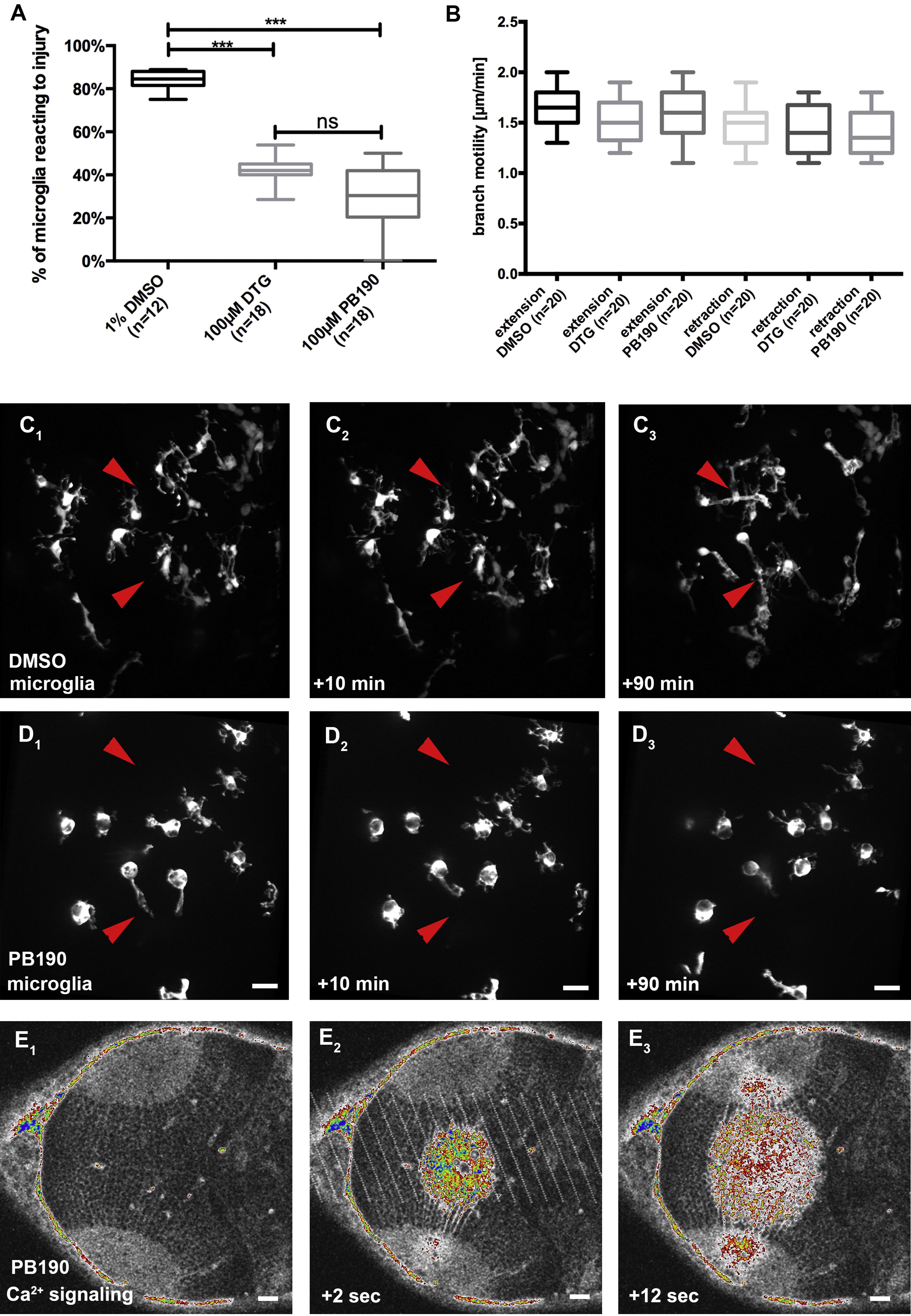
σ1 Receptor is important for microglial reaction toward injury.
(A) Percentage of microglia reacting to injury in DMSO compared to DTG and PB190 treated zebrafish larvae. (B) Speed of microglia branch extension and retraction in DMSO, DTG and PB190 treated embryos. (C) Time course of microglia (pU1::Gal4UAS::TagRFP) reaction toward injuries in DMSO treated embryos. (C1) before, (C2) 10 min after and (C3) 90 min after cut. (D) Time course of microglia (pU1::Gal4UAS::TagRFP) reaction toward injuries in PB190 treated embryos. (D1) before, (D2) 10 min after and (D3) 90 min after cut. (E) Dorsal views of a 3,5 dpf larval brain showing the time course of a Ca2+ wave (betaactin::GCaMP3.1) forming upon central brain injury in PB190 treated zebrafish larvae. (E1) before, (E2) 2 s after and (E3) 12 s after cut. Scalebar for all images 20 µm. Images (C) and (D) were obtained using an Andor Spinning Disk Confocal with a 40x/NA1.15 objective. Images (E) were produced using an Olympus FV 1000 with a 40x/NA1.15 objective. Statistical analysis was performed using a KruskalWallis test with a Dunn’s multiple comparison test.