
|
Fig. 1
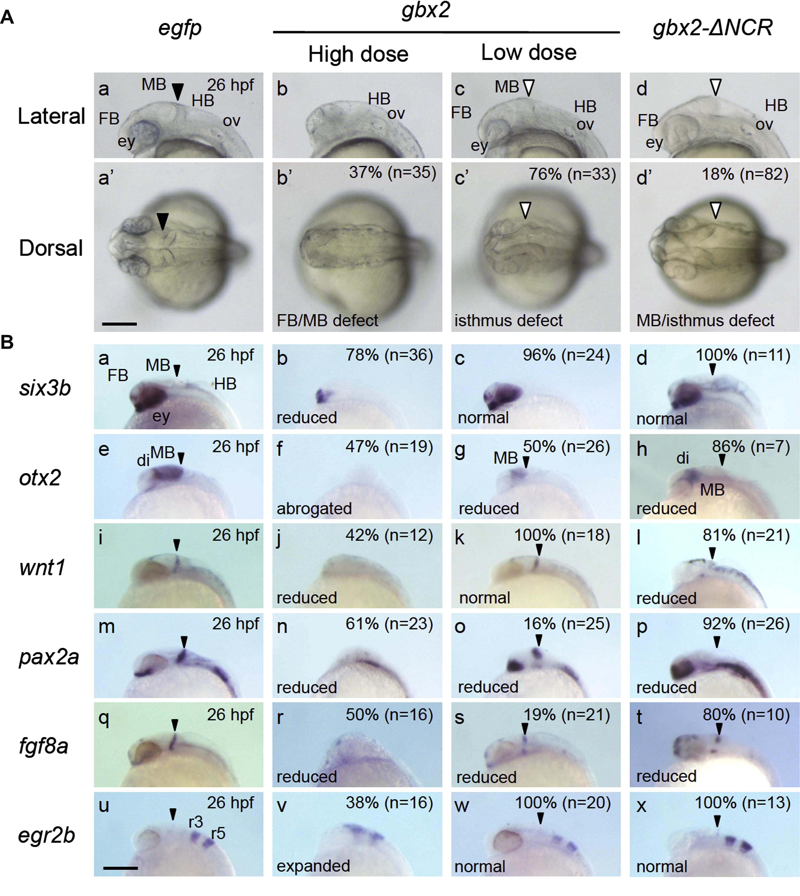
Effects of overexpression of gbx2 and gbx2-ΔNCR on brain development. Embryos were injected with gbx2 mRNA of 5 pg/embryo (low dose) or 30 pg/embryo (high dose) and gbx2-ΔNCR mRNA of 100 pg/embryo and examined at 26 hpf. (A) Head regions of embryos injected with mRNA. Lateral and dorsal views are shown in the top and bottom rows, respectively. di, diencephalon; ey, eye; FB, forebrain; HB, hindbrain; MB, midbrain; ov, otic vesicle. Normal and disrupted isthmuses are marked with solid and open large triangles, respectively. (B) Embryos injected with mRNA were examined at 26 hpf for the expression of brain regional markers shown on the left. Since the activities of gbx2 differed among mRNA preparations, the mRNA used in (A) was also injected throughout the series of experiments shown here. The most striking phenotypes and their proportions are shown in the respective panels. The positions of the MHB are marked with small triangles. Scale bars, 200 µm.
Reprinted from Mechanisms of Development, 130(11-12), Nakayama, Y., Kikuta, H., Kanai, M., Yoshikawa, K., Kawamura, A., Kobayashi, K., Wang, Z., Khan, A., Kawakami, K., and Yamasu, K., Gbx2 functions as a transcriptional repressor to regulate the specification and morphogenesis of the mid-hindbrain junction in a dosage- and stage-dependent manner, 532-52, Copyright (2013) with permission from Elsevier. Full text @ Mech. Dev.