
|
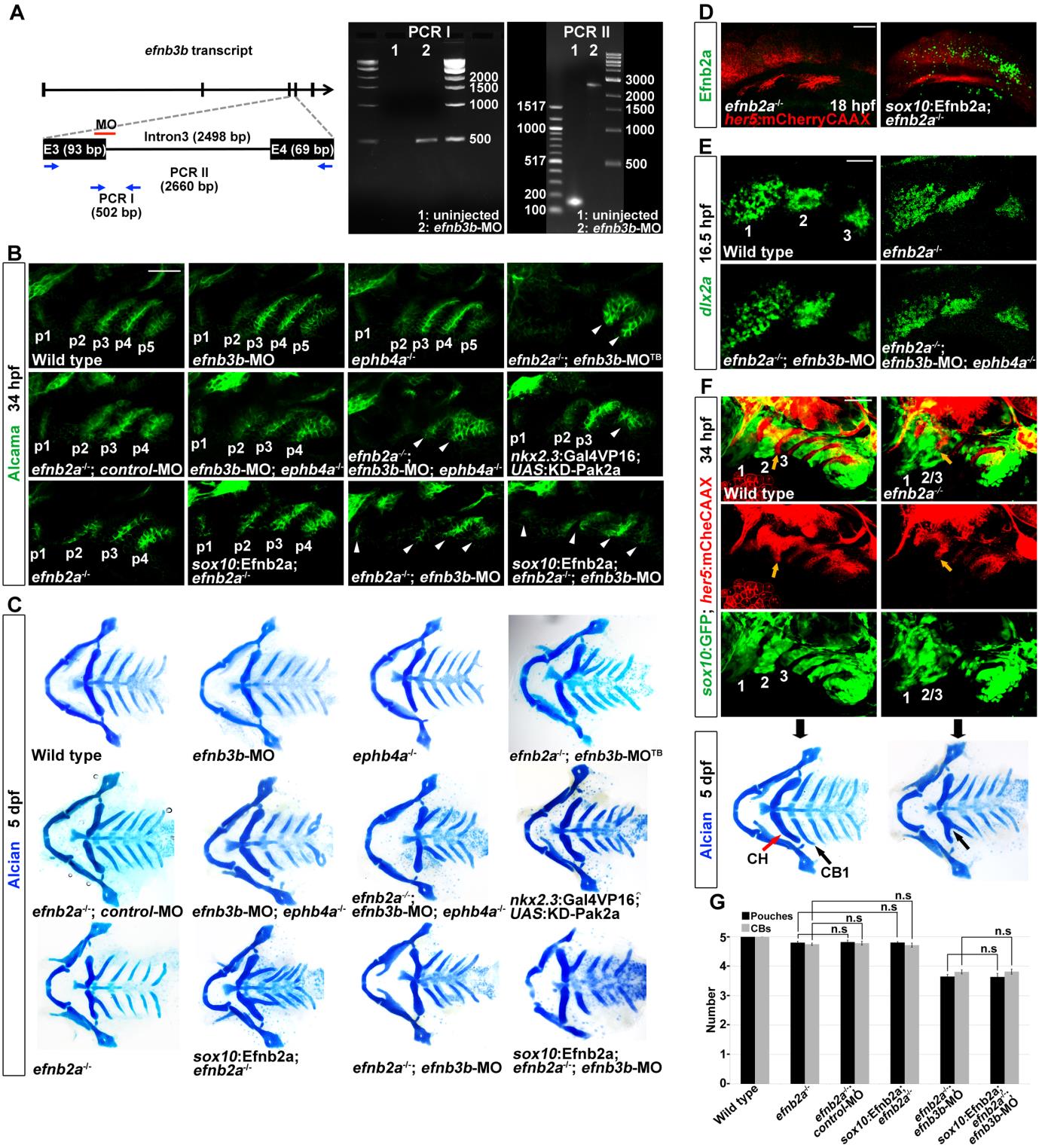
Fig. S2 EphB signaling in pouch, CB cartilage and neural crest development. (A) An MO was designed against the exon 3 (E3)-intron 3 splice junction of efnb3b. In uninjected control embryos, normal splicing of exons 3/4 resulted in excision of intron 3 (2498 bp) and loss of PCR amplification of 24-hpf embryonic cDNA by the designated primers (arrows of PCR I) and amplification of a 162 bp band spanning exons 3/4 (arrows of PCR II). By contrast, 24-hpf embryos injected with 1 nL of 300 µM efnb3b MO at the one-cell stage resulted in amplification of a 502 bp band (PCR I) and a 2660 bp band (PCR II), and loss of the 162 bp band spanning exons 3/4 (PCR II), due to a failure to splice out intron 3. (B) Alcama immunohistochemistry reveals normal pouches in efnb3b- MO and ephb4a-/- single mutants, and disrupted pouches in efnb3b-MO; ephb4a-/- and efnb2a-/-; efnb3b-MO; ephb4a-/- compound mutants. Whereas injection of a translationblocking efnb3b morpholino (efnb3b-MOTB) enhanced the defects of efnb2a mutant pouches, injection of a control morpholino antisense to the splice-blocking morpholino failed to enhance. Forced expression of a dominant-negative KD-Pak2a transgene in the nkx2.3+ endoderm (nkx2.3:Gal4VP16; UAS:KD-Pak2a) also resulted in reduced numbers of dysmorphic pouches. Compared with non-transgenic efnb2a-/- or efnb2a-/-; efnb3b-MO siblings, the sox10:Efnb2a transgene did not rescue pouches in EphrinB-deficient mutants. Arrowheads denote malformed pouches. Scale bar: 40 µm. (C) Alcian Blue staining of cartilage reveals no CB defects in efnb3b-MO and ephb4a-/- single mutants, and lost and/or fused CBs in efnb3b-MO; ephb4a-/- and efnb2a-/-; efnb3b-MO; ephb4a-/- compound mutants, as well as in embryos with endoderm-specific Pak2a disruption. Injection of a translation-blocking but not a control efnb3b-MO into efnb2a mutants enhanced CB defects of efnb2a mutants. sox10:Efnb2a transgene in either efnb2a single mutants or efnb2a-/-; efnb3b-MO compound animals failed to rescue losses of CBs and fusions of CH and CB1. (D) Immunohistochemistry at 18 hpf shows that Efnb2a protein is lost in efnb2a mutants and restored to migratory neural crest mesenchyme by the sox10:Efnb2a transgene. her5:mCherryCAAX labels pre-pouch endoderm where Efnb2a expression is not restored. Confocal projections are shown. Scale bar: 40 µm. (E) Fluorescent in situ hybridization for dlx2a (green) at 16.5 hpf shows no intermingling of neural crest cells between the three migrating streams (numbered) in embryos with losses of EphrinB ligands and EphB receptors. Scale bar: 40 µm. (F) Development of pharyngeal arches, pouches and cartilages in individual efnb2a mutants and wild-type siblings. In wild types at 34 hpf, labeling of neural crest-derived cells by sox10:GFP (green) shows six arches, and labeling by her5:mCherryCAAX (red) shows five pouches. Note that arches 2 and 3 are fully separated by the second pouch (yellow arrow), and by 5 dpf, Alcian Blue staining reveals that the CH and CB1 cartilages are well separated. In the efnb2a-/- example, the second pouch does not extend as far ventrally (yellow arrow) and arches 2 and 3 appear fused ventrally. In this same animal at 5 dpf, CH and CB1 cartilages are also fused ventrally (arrow). Scale bar: 40 µm. (G) Quantification of pouch and CB defects. Data represent mean±s.e.m. n.s., not significant.