
|
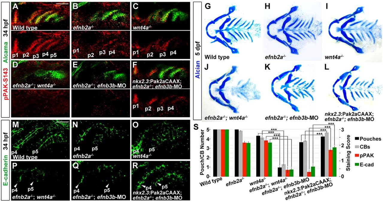
Fig. 4
Requirements for Eph-ephrin and Wnt signaling in PAK activation and E-cadherin levels. (A-F) Immunohistochemistry shows Alcama (green) and the activated form of PAK (pPAK-S143, red) at 34hpf. In wild types, pPAK-S143 is higher in anterior pouches (p1-p3) compared with less mature posterior pouches (p4 and in particular p5). Compound efnb2a; wnt4a mutants produce fewer pouches and have further reduced pPAK-S143 staining than single mutants alone. In EphrinB-deficient embryos, forced expression of membrane-targeted Pak2a (nkx2.3:Pak2aCAAX) rescues both pouches and PAK autophosphorylation. Scale bar: 40µm. (G-L) Ventral views of facial cartilage at 5dpf show losses of CBs and CH-CB1 fusions in mutants and rescue of these defects in EphrinB-deficient embryos by nkx2.3:Pak2aCAAX. (M-R) Immunohistochemistry shows E-cadherin protein (green) within pouches p4 and p5. In wild types, E-cadherin localizes to cell-cell contacts along apicolateral membranes. Compound efnb2a; wnt4a mutants have further reduced E-cadherin staining than single mutants alone, and nkx2.3:Pak2aCAAX rescues E-cadherin levels in EphrinB-deficient embryos. Scale bar: 20µm. (S) Quantification of pouch and CB defects, and scoring of reduced pPAK and E-cadherin staining. Data represent mean±s.e.m. ***P<0.001.