
|
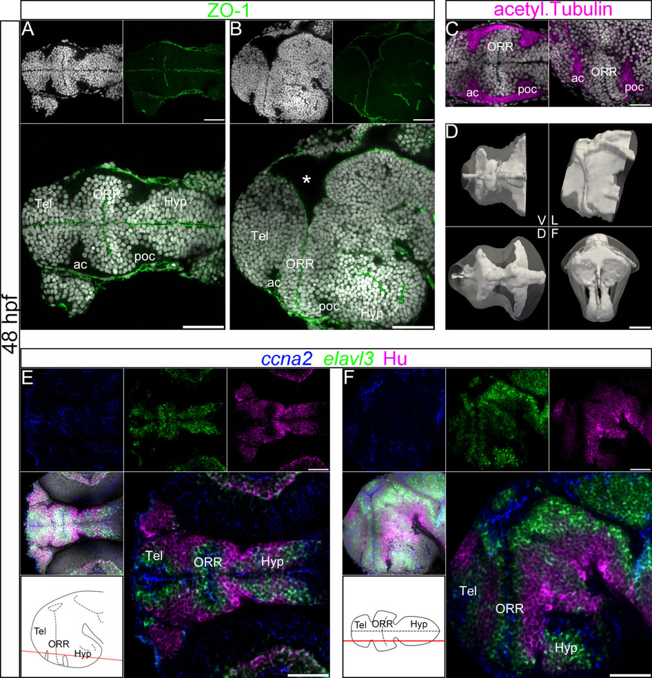
Fig. 1
Ventricular organization and the centrifugal gradient of neurogenesis at 48 hpf.
(A–C): Single confocal plane of a 48hpf embryonic forebrain stained with DAPI (gray), immunolabeled for ZO-1 (A and B) or acetylated α-tubulin (C), in ventral (A and left panel in C) and lateral (B and right panel in C) views. The telencephalon (Tel), the optic recess region (ORR), and the hypothalamus (Hyp) form three distinct cellular regions in the secondary prosencephalon. The ORR is bordered by the dense fiber bundles, the anterior commissure (ac) and post-optic commissure (poc). The asterisk (*) in (B) (lateral view) indicates the enlargement of the dorsal ventricular lumen corresponding to the anterior intraencephalic sulcus (AIS). (D): Surface rendering of the shape of ventricle (white, in opacity), reconstructed from ZO-1 immunolabeling, overlapped on the shape of the brain (white, in transparency), reconstructed from DAPI staining. V = ventral, D = dorsal, L = lateral and F = frontal views. The 3D representation of both ventricle and brain facilitates the visualization of the convoluted ventricular organization in the forebrain. (E–F): Single confocal plane of a 48hpf embryonic forebrain labeled for the neurogenic markers ccna2, elavl3 and HuC/D in ventral (E) and lateral (F) views. Bottom left drawings show the level of corresponding optical planes. ccna2-positive proliferative cells are concentrated around the ventricular zones, while elavl3-positive differentiating and HuC/D-positive differentiated cells are located at the periphery of the neural tube. Scale bars = 50µm.