
Fig. 3
- ID
- ZDB-IMAGE-150422-3
- Genes
- Publication
- Marjoram et al., 2015 - Epigenetic control of intestinal barrier function and inflammation in zebrafish
- All Figures
- Figures for Marjoram et al., 2015
|
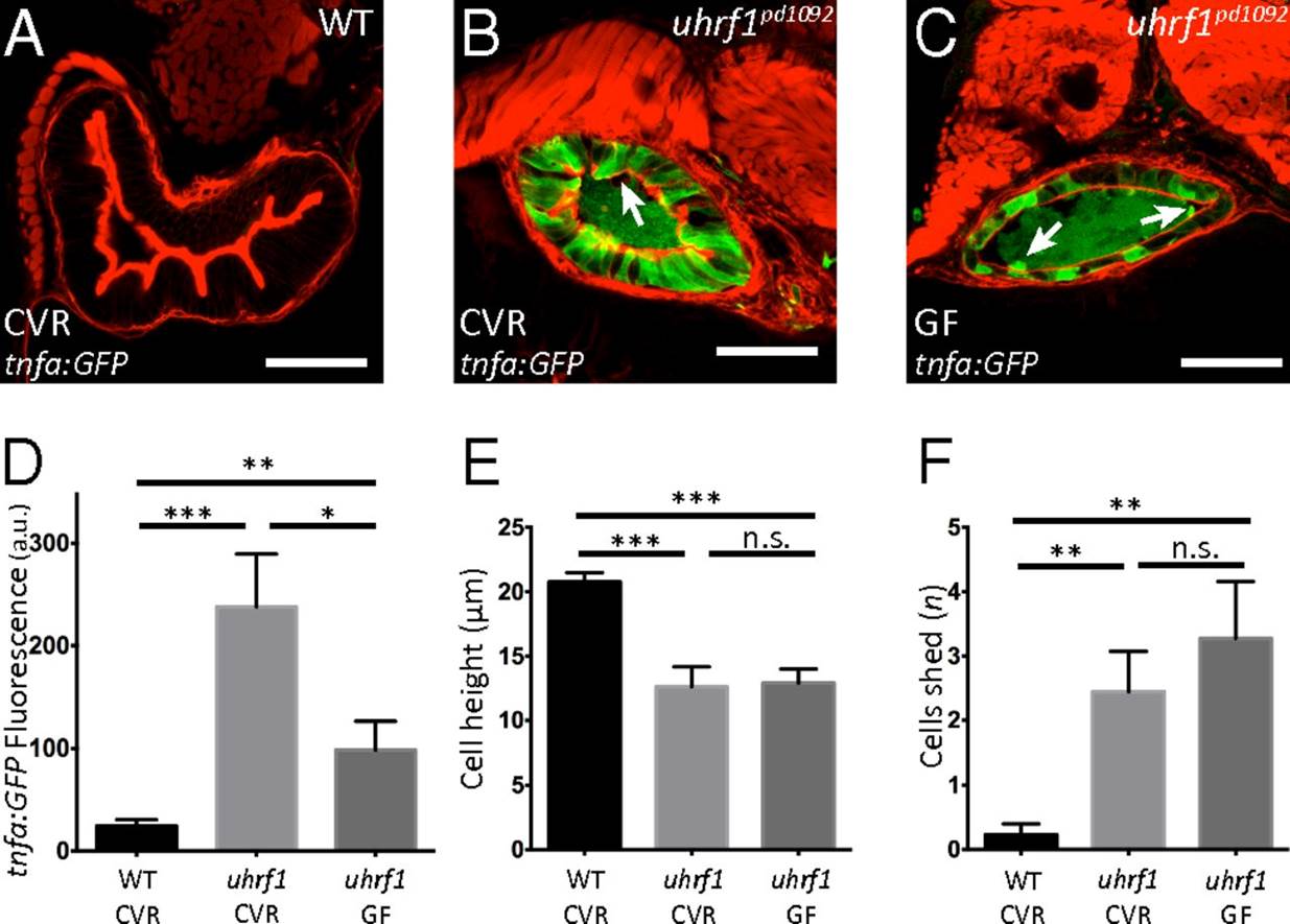
Fig. 3
Microbiota presence enhances tnfa elevation in the uhrf1pd1092 gut epithelium but is not required for the development of an IBD-like phenotype. (A–C) Confocal images of cross-sections of conventionally raised (CVR) 120 hpf WT (A); CVR 120 hpf uhrf1pd1092 (B), and germ-free (GF) 120 hpf uhrf1pd1092 (C) larvae. The arrows in B and C mark cells being shed from the epithelium. (D) TgBAC(tnfa:GFP) fluorescence (measured in arbitrary units, a.u.) under CVR and GF conditions. (E) Cell height measurement under CVR and GF conditions. Cell height is unchanged between CVR and GF mutants. (F) Quantification of cell shedding. Cell shedding was not decreased in GF mutants. Phalloidin, red. (Scale bars: 50 µm.) (D–F: n = 13 WT CVR; n = 9 mutant CVR; n = 11 mutant GF). Bars represent mean ± SEM. Significant differences between the means were detected after an ANOVA. *P < 0.05, **P < 0.01, ***P < 0.0001, n.s., not significant. Show Less