
|
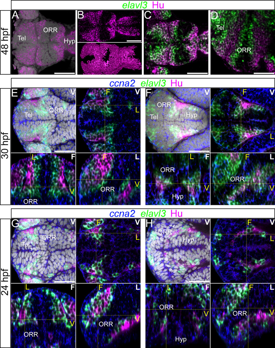
Fig. 4
Differentiated cells mark the boundaries of each morphogenetic entity of the secondary prosencephalon.
(A–D): 48hpf forebrain labeled for elavl3 and HuC/D superimposed on DAPI staining (gray). (A–B): HuC/D labeling with DAPI (A) and without DAPI (B). The top panel of B shows a single confocal plane of a ventral view and bottom panel of B shows the surface rendering of segmented HuC/D immunolabeling alone. These images show that the HuC/D staining itself is able to recapitulate the outline of the three morphogenetic entities. (C–D): Single confocal plane of a ventral (C) and lateral (D) view of elavl3 and Hu labeling. Two rows of HuC/D-positive cells are in apposition at boundaries: the boundary between the telencephalon (Tel) and the optic recess region (ORR), and the boundary between ORR and the hypothalamus (Hyp). (E–H): Single confocal plane of the forebrain labeled with the neurogenic markers ccna2, elavl3 and HuC/D at 30hpf (E–F) or 24hpf (G–H) in different views. In each case, the ventral view (indicated “V” in white) is shown on the top (with DAPI on the left, without DAPI on the right). The bottom images show the frontal (indicated “F” in white) and lateral (indicated “L” in white) views reconstructed using Z-projections of the ventral images. As they are ventral, frontal and lateral views from a single embryo, corresponding section levels of different views are indicated in yellow lines and yellow letters. As shown at 48hpf (A–D), two layers of HuC/D labeling are detected around the boundary of the three cellular masses however this is not visible at the same section level. E and G show HuC/D and elavl3 expression at the telencephalic/ORR border whereas F and H show at the ORR/hypothalamic region border. Scale bars = 50µm.