
Image
|
Figure Caption
Fig. 6, S4
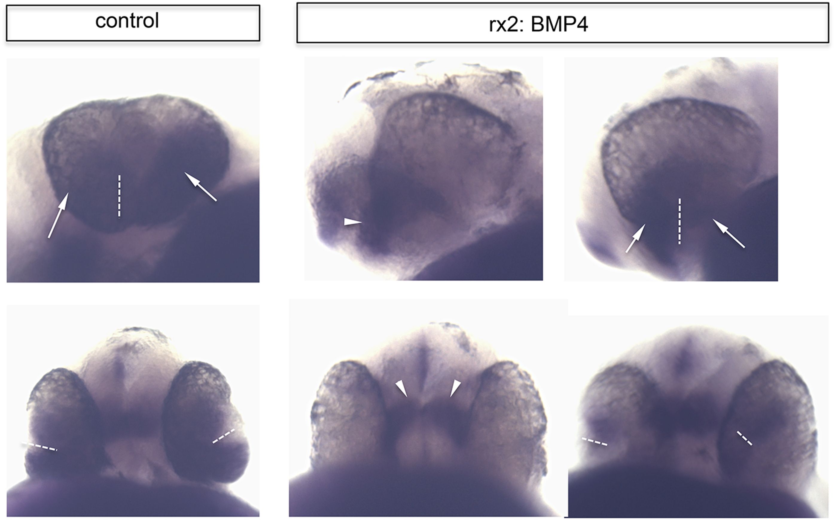
Ventral retinal identity remains in rx2::BMP4 embryos.
Whole mount in situ hybridization with a vax2 probe (NBT/BCIP) of control (left) and rx2::BMP4 embryos (right) in a lateral view (upper pictures) and a ventral view (lower pictures) (28 hpf). Note that the ventral retinal marker remains expressed in the forming ventral optic cup even if BMP4 is expressed panocularly (rx2::BMP4). Also note that the vax2 domain in control is broader than in the rx2::BMP4 embryos (arrows) in which it is more prominent in the optic stalk region (arrowhead) (the dotted line indicates the optic fissure).
Acknowledgments
This image is the copyrighted work of the attributed author or publisher, and
ZFIN has permission only to display this image to its users.
Additional permissions should be obtained from the applicable author or publisher of the image.
Full text @ Elife