
|
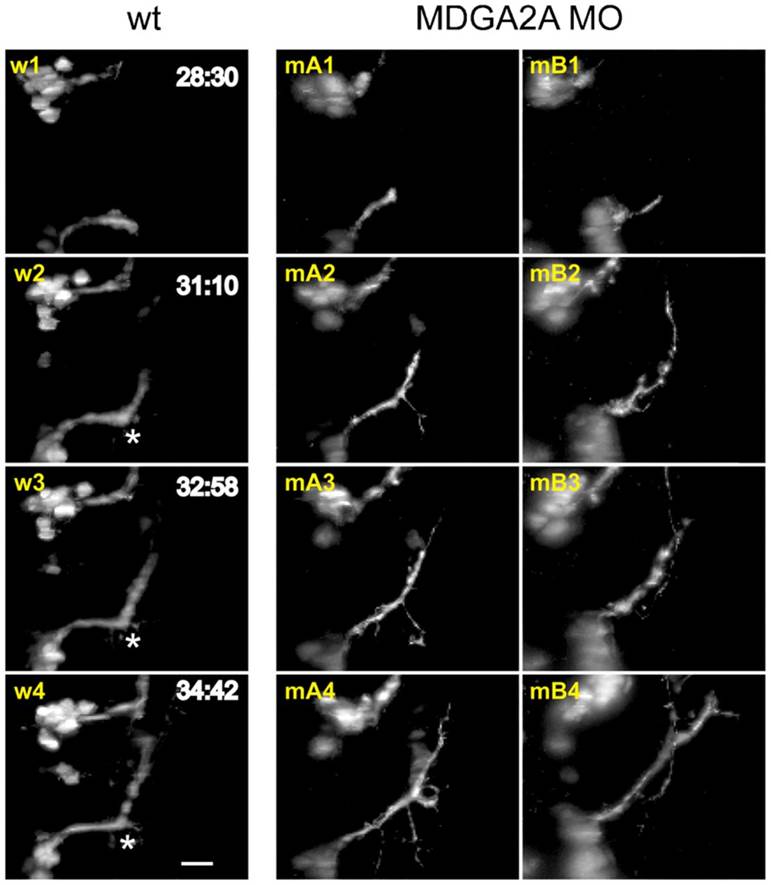
Fig. 5
Representative images from control (w) and MDGA2A morpholino treated (mA and mB) embryos are shown. 28 to 34h post fertilization facial neurons in wt embryos project axons along a predetermined path, displaying a well-documented 60° turn (asterisks). In MDGA2A morphants this turning angle is absent or strongly reduced and defasciculation events occur much more frequently. Analyzing fluorescence intensity below the main axon bundle of the facial nerve indicates that fluorescence in MDGA2A knockdown embryos is slightly increased, supporting the finding of increased defasciculation and axon branching of facial neurons (see supplementary material Fig. S3D–F). The scale bar represents 20µm.