
|
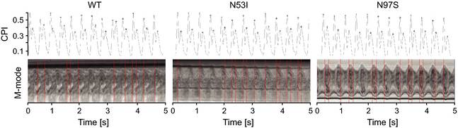
Fig. 3
Monitoring of embryo heart contractions. Examples of changing pixel intensities (CPIs) and M-modes for β-adrenergic stimulated embryos injected with CaM WT, N53I or N97S mRNA, respectively. CPIs have peaks at the onset of diastole and systole (stars). M-mode shows embryonic ventricle movement. The mean diastolic interval was 0.173 ± 0.028 s for the control and 0.177 ± 0.017 s for CaM WT compared to 0.156 ± 0.023 s and 0.151 ± 0.025 s for CaM N53I and for CaM N97S (both P < 0.05 compared to WT), respectively. The mean systolic interval was 0.264 ± 0.024 s for the control and 0.263 ± 0.027 CaM WT compared to 0.256 ± 0.031 s for CaM N53I and 0.262 ± 0.027 for CaM N97S. No significant difference in systolic interval was observed (P = 0.9).