
|
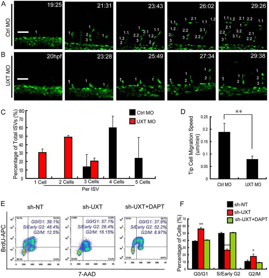
Fig. 4
UXT modulates endothelial cell migration and cell cycle. (A,B) Confocal time-lapse images (from 19hpf to 30hpf) of Tg(kdrl:EGFP)s843 embryos injected with 4ng of control MO (A) or 4ng of UXT MO (B). The numbering on the images shows the indicated time points. Scale bars: 50µm. (C) Quantification of endothelial cell number in the segmental artery sprouts. (D) Quantification of the migration speed of ISV tip cells in control embryos and UXT morphants. (E,F) The inhibitory effect of UXT on cell cycle. (E) Flow cytometric analysis for BrdU incorporation and 7-AAD labeling in wild-type and UXT-deficient HUVECs or UXT-deficient HUVECs treated with 1.5µM DAPT. (F) Quantification of the percentage of cells in different cell cycle stages. All quantitative data are the mean±s.e.m. (at least three independent experiments);*P<0.05, **P<0.01, ***P<0.001 versus the corresponding control.