
|
Fig. S4
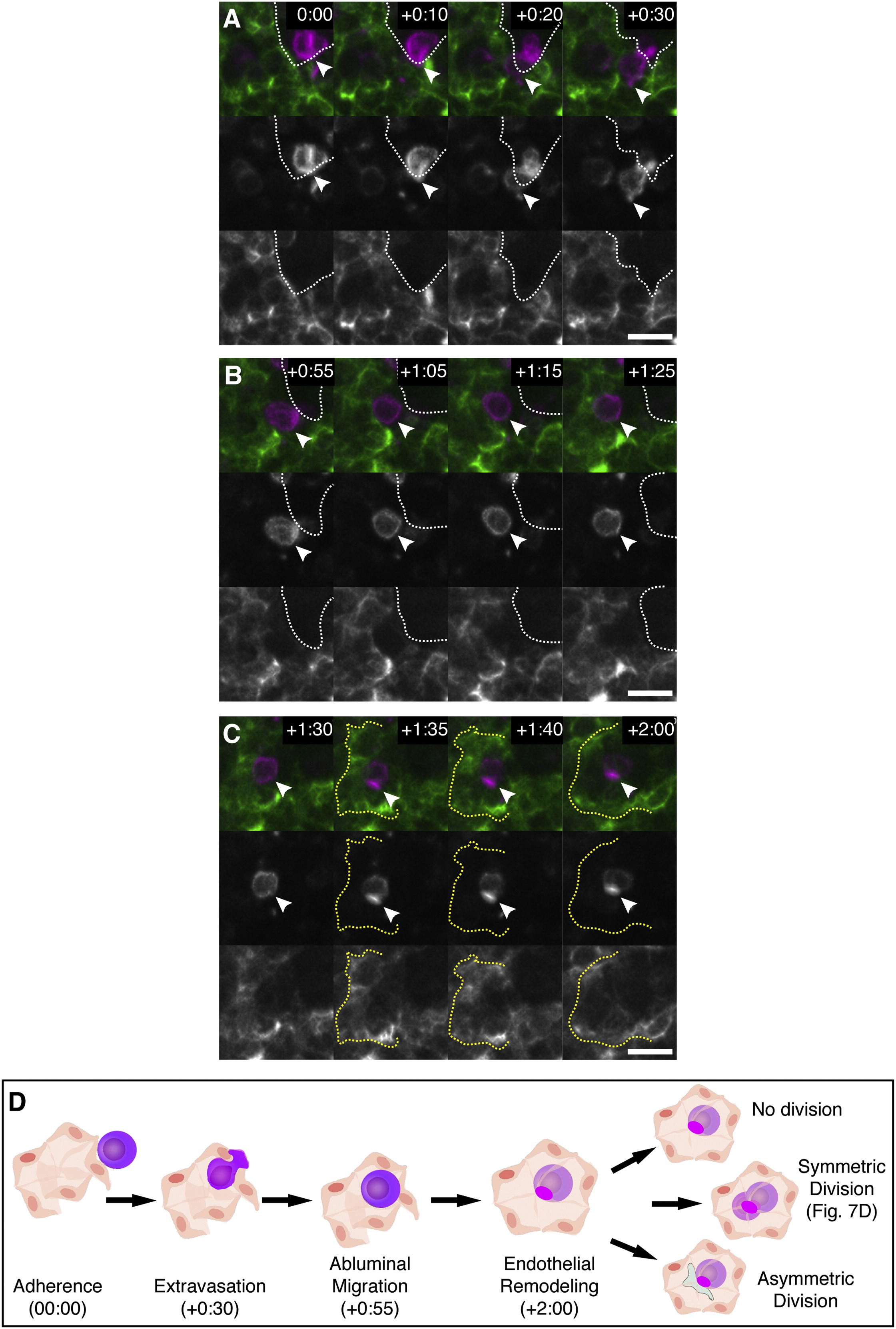
Time-Lapse Live Imaging of E11.5 FL Explant with Labeled HSPC and ECs, Related to Figure 4
(A) A time-lapse sequence from a FL lobe showing a c-kit+ HSPC (magenta) adhered to a CD31+ sinusoid (green). In ~30 min, the anterior of the cell protrudes (arrowhead) and extravasates to the abluminal side of the sinusoid (boundary marked by white dotted line). Luminal versus abluminal sides of the sinusoid were confirmed in single z slices of confocal stacks (data not shown). Times are hours:minutes after start of explant imaging. Scale bars: 10 µm.
(B) After extravasation, the HSPC migrates on the abluminal side of the sinusoid.
(C) ECs surround the HSPC as a rosette (yellow dotted line).
(D) A diagram summarizing the steps of HSPC lodgement in the FL. The times shown correspond to the time-lapse frames shown above. A morphologically symmetric division is observed in Figure 4D. Other possible cell division decisions are shown.
Reprinted from Cell, 160, Tamplin, O.J., Durand, E.M., Carr, L.A., Childs, S.J., Hagedorn, E.J., Li, P., Yzaguirre, A.D., Speck, N.A., Zon, L.I., Hematopoietic Stem Cell Arrival Triggers Dynamic Remodeling of the Perivascular Niche, 241-252, Copyright (2015) with permission from Elsevier. Full text @ Cell