
|
Fig. S3
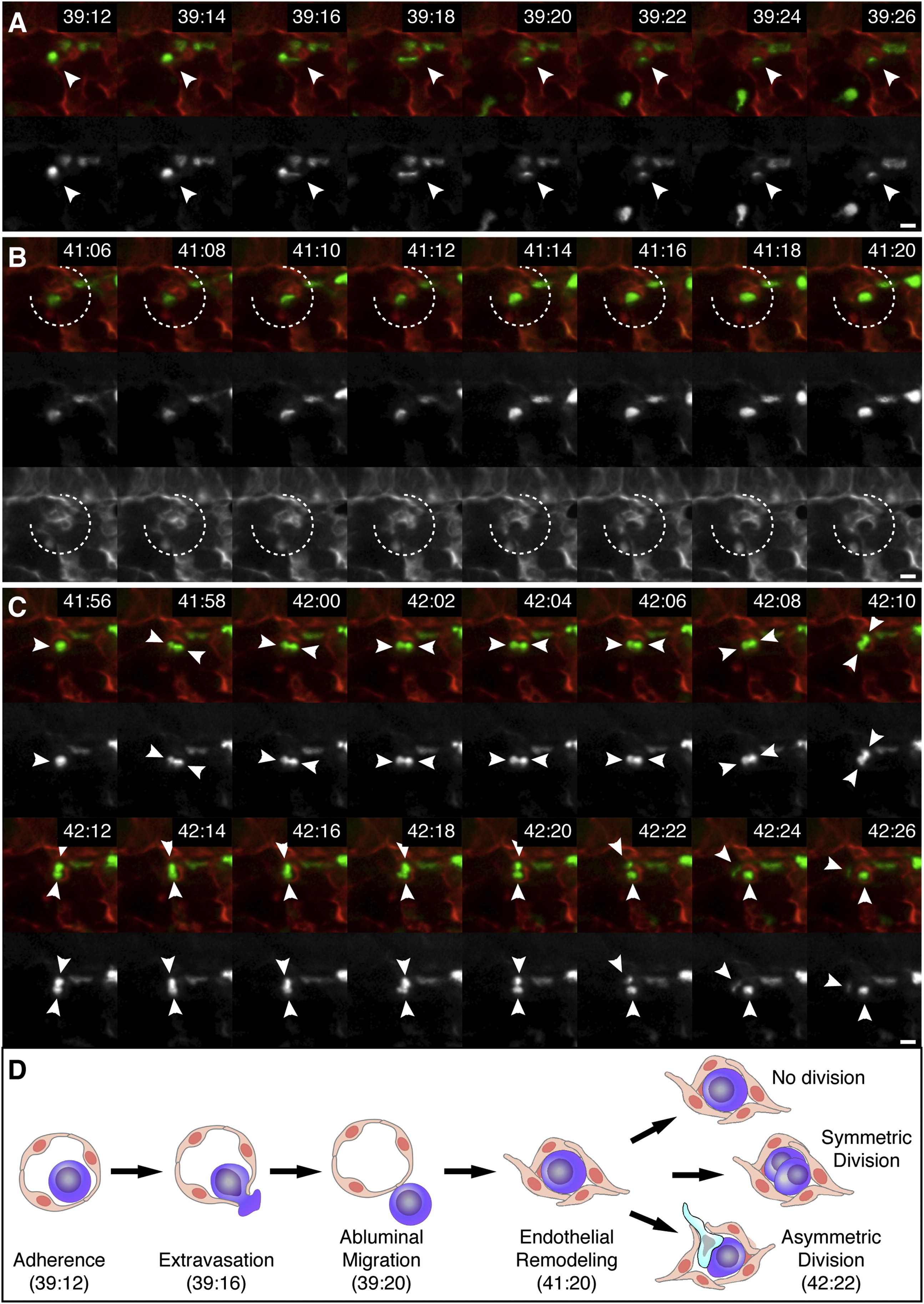
Time-Lapse Live-Imaging Sequence of CHT Colonization, Related to Figure 3 and Movie S2
Selected frames of time-lapse are shown (1 frame/2 min). First row frames are a merge of Runx:GFP+ HSPCs (green; second row) and kdrl:RFP+ ECs (red; third row in B). Times are hours:minutes post fertilization. Scale bars: 10 µm.
(A) HSPC (arrowhead) extravasates by squeezing through endothelial wall.
(B) ECs remodel around HSPC to form niche (broken circle).
(C) After HSPC division the daughter cells undergo a 90° rotation (arrowheads). The upper cell becomes migratory and crawls out of niche.
(D) A diagram summarizing the steps of HSPC lodgement in the perivascular niche. The times shown correspond to the time-lapse frames shown above. Other possible cell division decisions are shown.
Reprinted from Cell, 160, Tamplin, O.J., Durand, E.M., Carr, L.A., Childs, S.J., Hagedorn, E.J., Li, P., Yzaguirre, A.D., Speck, N.A., Zon, L.I., Hematopoietic Stem Cell Arrival Triggers Dynamic Remodeling of the Perivascular Niche, 241-252, Copyright (2015) with permission from Elsevier. Full text @ Cell