
|
Fig. 5
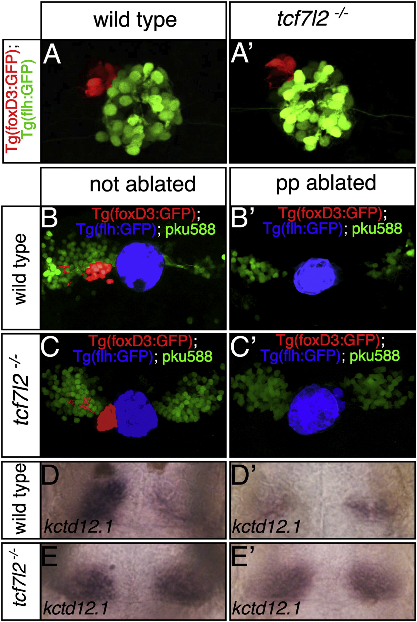
The Symmetric dHb Phenotype of tcf7l2 Mutants Is Epistatic to Parapineal Ablation
Dorsal views focused on the epithalamus of 2.5 dpf (A and A′) and 4 dpf (B–E′) embryos with anterior to the top.
(A and A′) The parapineal (pseudocolored red) is on the left in both wild-type and tcf7l2 mutant embryos.
(B–E′) In Et(gata2a:EGFP)pku588; Tg(foxD3:GFP); Tg(flh:eGFP) triple-transgenic normal and parapineal-ablated embryos, dHbl neurons (green) and the pineal complex are labeled. The pineal is pseudocolored in blue and the parapineal and its projections in red (B–C′). Embryos as in (B)–(C′) were labeled for kctd12.1 subsequent to live imaging (D–E′).