
|
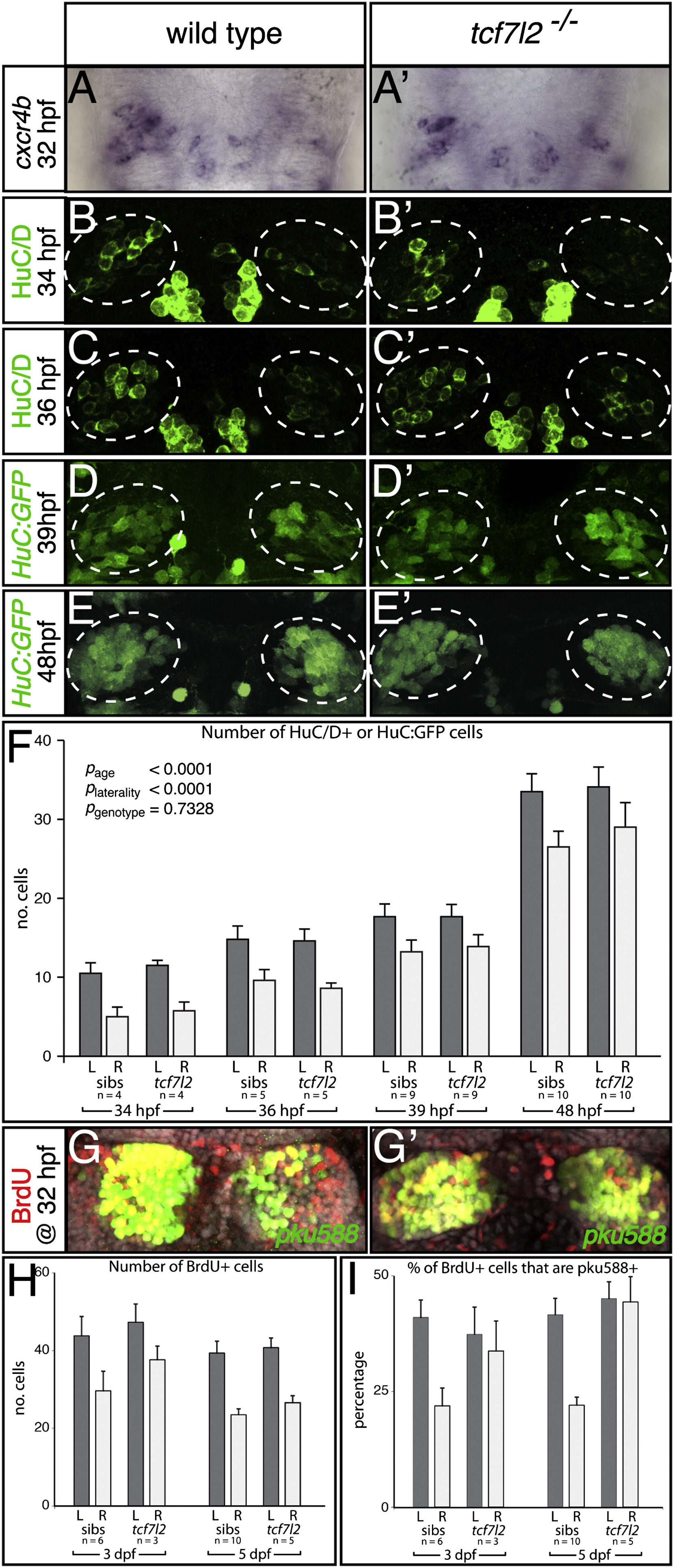
Fig. 3
Habenular Neurogenesis Is Not Overtly Affected in tcf7l2 Mutants
(A–E′, G, and G′) Dorsal views of the epithalamus, with anterior to the top.
(A and A′) The numbers and LR asymmetry of cxcr4b-expressing dHb neuronal precursors is not overtly affected in the tcf7l2 mutant.
(B–E′) HuC/D (B–C′) and transgenic HuC:GFP (D–E′) expression in differentiating habenular neurons in wild-type fry and tcf7l2 mutants. The dotted circles highlight the dHb.
(F) Numbers of HuC/D+ and HuC:GFP+ neurons are not significantly different between wild-type embryos and tcf7l2 mutants at 34, 36, 39, and 48 hpf. Error bars indicate the SEM.
(G and G′) BrdU (red)-labeled, 5 dpf dHb neurons expressing the pku588 transgene (green) in a wild-type (G) and a tcf7l2 mutant (G′).
(H and I) Graphs representing the number of BrdU-labeled cells (H) and the percentage of BrdU-labeled cells coexpressing the pku588 transgene (I) in wild-type embryos and tcf7l2 mutants. Error bars indicate the SD.
See also Table S2.