
|
Fig. 6
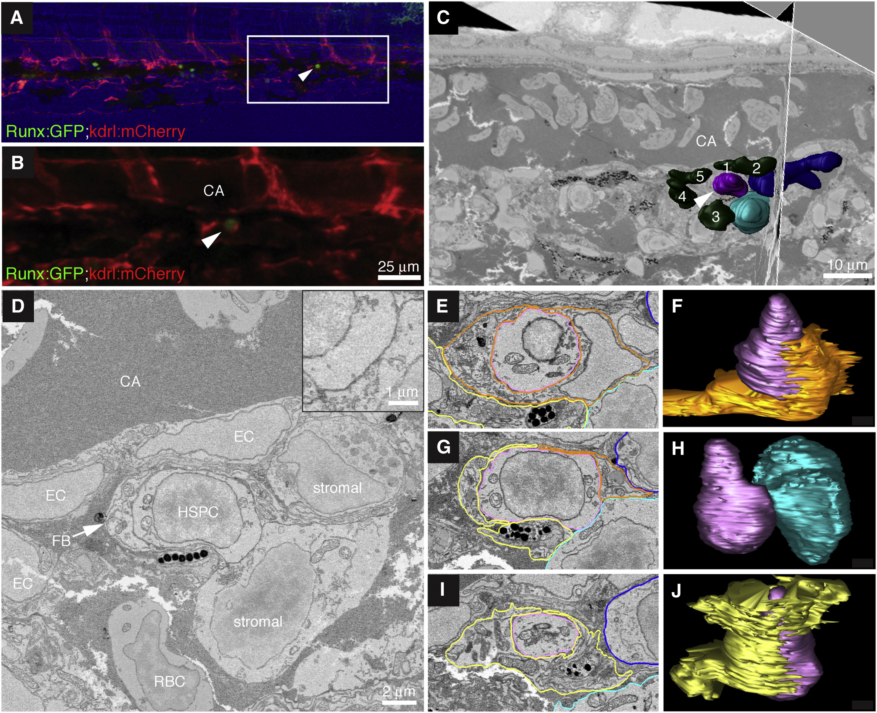
High-Resolution Electron Microscopy of Endogenous HSPC in the Perivascular Niche
(A) Last frame of CHT time lapse (60 hpf). Arrowhead marks HSPC lodged >6 hr. Runx:GFP (green), kdrl:mCherry (red), bright field (blue). Anterior left, posterior right, dorsal top, ventral bottom.
(B) Detail of region in (A) marked by box.
(C) Single section and orthogonal slice from serial block face EM scans. Lodged HSPC (purple, arrowhead), surrounding EC nuclei (green, numbered), and stromal cells (dark and light blue).
(D) High-resolution EM of HSPC lodged in perivascular niche ventral to DA. The HSPC is in direct contact with one stromal cell (see higher-magnification inset).
(E–J) Selected sections (left) through niche with cell membrane traces used to build 3D models (right).
(E) In this section, the HSPC (purple) is mostly surrounded by EC (orange). Portions of fibroblastic (yellow) and stromal (light and dark blue) cells are visible.
(F) About half of the HSPC surface is wrapped by EC.
(G) The HSPC directly contacts the stromal cell. Portions of the fibroblastic cell, EC, and second stromal cell are visible.
(H) Only the midsection of the HSPC contacts the stromal cell.
(I) The fibroblastic cell surrounds the HSPC. Portions of two stromal cells are visible.
(J) Most of the HSPC surface is wrapped by the fibroblastic cell.
Scale bars, (B) 25 µm; (C) 10 µm; (D) 2 µm; and inset, 1 µm. See also Figure S5 and Movie S7.
Reprinted from Cell, 160, Tamplin, O.J., Durand, E.M., Carr, L.A., Childs, S.J., Hagedorn, E.J., Li, P., Yzaguirre, A.D., Speck, N.A., Zon, L.I., Hematopoietic Stem Cell Arrival Triggers Dynamic Remodeling of the Perivascular Niche, 241-252, Copyright (2015) with permission from Elsevier. Full text @ Cell