
|
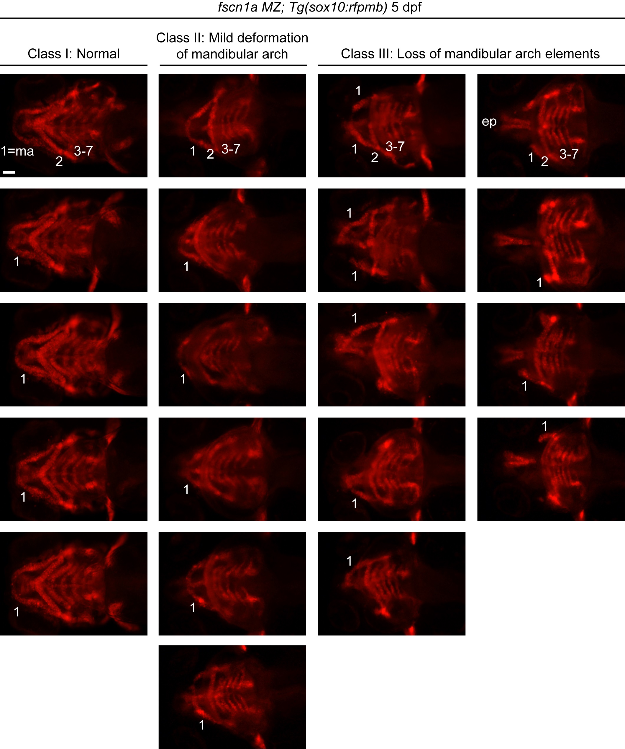
Fig. S7 fscn1a MZ embryos display abnormal craniofacial skeleton morphology.
Representative images of craniofacial skeleton phenotypes observed in fscn1a MZ embryos. Ventral views of craniofacial skeleton in 5 dpf Tg(sox10:rfpmb); fscn1a MZ embryos. Phenotypes are grouped into three classes: normal craniofacial cartilage morphology (Class I), mild deformation of mandibular arch (Class II) and loss of mandibular arch elements (Class III). Within a clutch of fscn1a MZ embryos, ~80% of embryos belong to Class I or II. The remaining 20% of embryos display symmetric or asymmetric loss of mandibular arch elements of variable severity and belong to Class III. Numbers denote pharyngeal arches, ma = mandibular arch (arch 1), ep = ethmoid plate. Scale bar = 100 µm.