
|
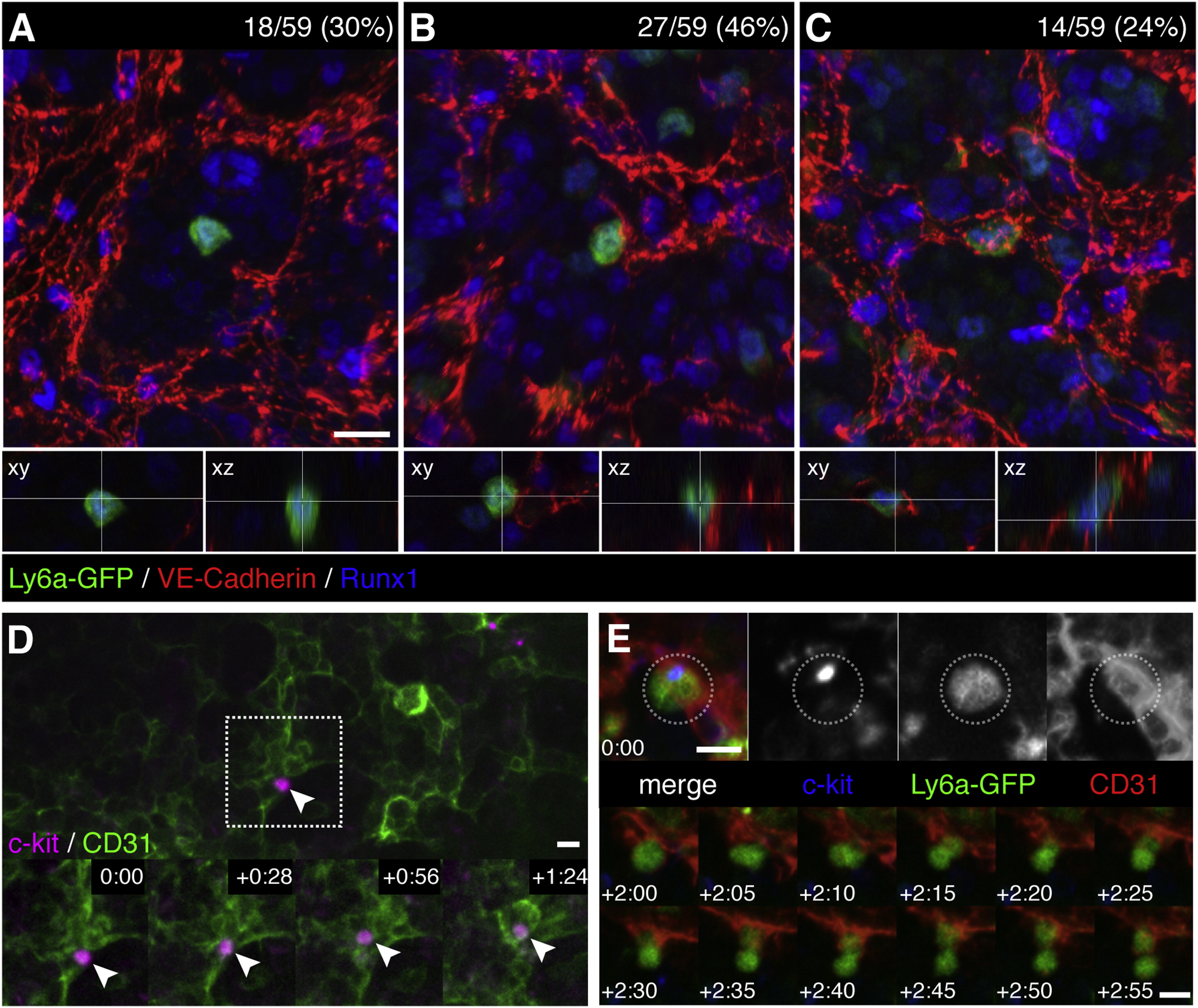
Fig. 4
Endothelial Cells Surround HSPCs in the Fetal Liver Microenvironment
(A–C) FLs from E11.5 Ly6a-GFP mice were fixed and stained for immunofluorescence with anti-VE-Cadherin (red), anti-Runx1 (blue), and anti-GFP (green) antibodies. We scored 59 Ly6a-GFP+/Runx1+ cells from 3 FLs and identified 3 different HSPC-EC configurations: (A) abluminal with no contact between HSPC and ECs (18/59; 30%); (B) EC contact on one side of the HSPC (27/59; 46%); and (C) HSPC surrounded on all sides with ECs (14/59; 24%). See Movie S3.
(D) c-kit+ cell (magenta) adhered to CD31+ ECs (green) in one lobe of an E11.5 FL (arrowhead). White box marks details below. Time-lapse frames show in <90 min the HSPC migrates into a field of ECs. Soon after, ECs surround HSPC to form niche. See also Movie S4.
(E) c-kit+(blue)/Ly6a-GFP+(green) HSPC adhered to abluminal side of CD31+ EC (red). Following this cell >2 hr (1 frame/5 min) shows a division with distal and proximal daughters relative to sinusoid; the latter remains in an endothelial surround. See also Movie S5. Confocal images: 3D rendered depth projection (A, B, C, and E), orthogonal view (A, B, and C below), maximum projection (D) of z stack.
Scale bars, 10 µm. See also Figure S4.
Reprinted from Cell, 160, Tamplin, O.J., Durand, E.M., Carr, L.A., Childs, S.J., Hagedorn, E.J., Li, P., Yzaguirre, A.D., Speck, N.A., Zon, L.I., Hematopoietic Stem Cell Arrival Triggers Dynamic Remodeling of the Perivascular Niche, 241-252, Copyright (2015) with permission from Elsevier. Full text @ Cell