
|
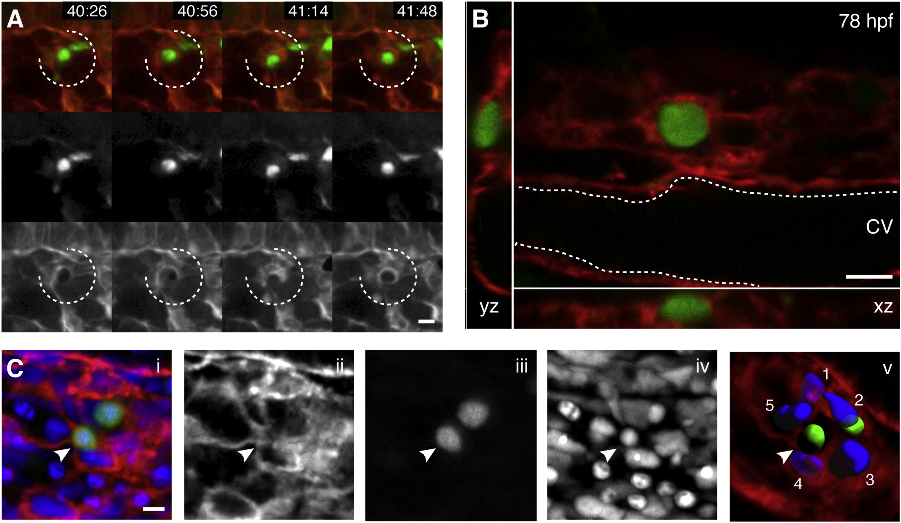
Fig. 3
Endothelial Cells in the Perivascular Niche Remodel to Surround a Single HSPC
(A) Four frames from time-lapse Movie S2 40–42 hpf (hr post fertilization:min). Upper row is a merge of Runx:GFP+ HSPC (green, middle row) and kdrl:RFP ECs (red, lower row). A group of surrounding ECs (broken circle) remodel around a single HSPC soon after its arrival.
(B) Higher magnification (60×) live image of single Runx:GFP+ HSPC surrounded by kdrl:mCherry ECs at 78 hpf (orthogonal views).
(C) 3D rendered projection of scanning confocal image from fixed 80 hpf embryo. (i) merge, (ii) kdrl:mCherry ECs, (iii) Runx:GFP+ HSPCs, (iv) DRAQ5 nuclei, and (v) kdrl:mCherry projection with 3D modeled green HSPCs and five blue surrounding EC nuclei (arrowhead indicates HSPC in EC surround).
All views: dorsal up, ventral down. Scale bars, 10 µm. See also Figure S3.
Reprinted from Cell, 160, Tamplin, O.J., Durand, E.M., Carr, L.A., Childs, S.J., Hagedorn, E.J., Li, P., Yzaguirre, A.D., Speck, N.A., Zon, L.I., Hematopoietic Stem Cell Arrival Triggers Dynamic Remodeling of the Perivascular Niche, 241-252, Copyright (2015) with permission from Elsevier. Full text @ Cell