
|
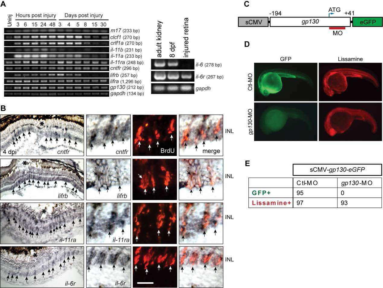
Fig. S5 Genes encoding IL-6 family cytokines are expressed in MG-derived progenitors upon retinal injury. Related to Figure 4. (A) RT-PCR analysis of retinal gene expression at various times after injury. il-6 is expressed in adult kidney and 8 dpf larva, but undetectable in the injured retina. (B) In situ hybridization shows induction of il-6 family member genes in BrdU+ MG-derived progenitors at the injury site. The asterisks mark the injury sites and the arrows point to MG-derived progenitors. Scale bars, 20 µm. (C) Diagram of sCMV:gp130-egfp reporter in which a fragment of gp130 DNA, containing the MO target sequence, is in-frame with the egfp coding sequence and under control of the sCMV promoter. (D) Injection of the sCMV:gp130-egfp reporter together with either lissamine-tagged control (Ctl) MO or gp130-targeting MO into one cell stage of zebrafish embryos and examined by fluorescence 24 hours later. Red fluorescence shows embryos received lissamine-tagged MOs. (E) Quantification of GFP-expressing embryos at 24 hpf. No GFP was detected in the gp130-targeting MO injected group, while GFP was readily detected in the control group. Similar results were obtained in 3 independent experiments. dpf, days post fertilization ; INL, inner nuclear layer.