
|
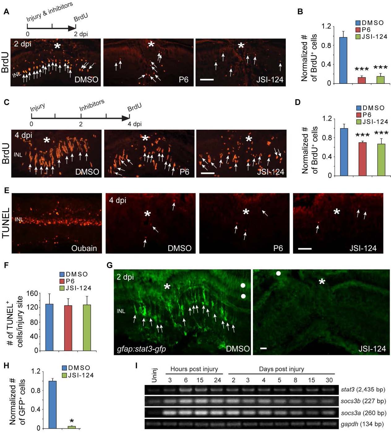
Fig. S3 Jak/Stat signaling regulates proliferation of MG-derived progenitors and Stat3-GFP expression. Related to Figure 2. (A, B) BrdU immunofluorescence on retinal sections shows that exposing fish to Jak inhibitors P6 or JSI-124 from 0-2 dpi inhibits progenitor proliferation at 2 dpi; ***P<0.001, n=4. (C, D) BrdU immunofluorescence shows that exposing fish to Jak inhibitors P6 or JSI-124 from 2-4 dpi inhibits progenitor proliferation at 4 dpi; ***P<0.001, n=4. (E, F) TUNEL staining for apoptotic cells at 4 dpi in retinas treated with DMSO, P6 or JSI-124. Oubain was used as a positive control. In panel F, n=3. (G, H) GFP immunofluorescence shows that exposing gfap:stat3-gfp fish to Jak inhibitors JSI-124 from 0-2 dpi inhibits Stat3-GFP expression; *P<0.05, n=3. (I) RT-PCR analysis of retinal stat3 and socs3 gene expression at various times after injury. Asterisks mark the injury sites (needle poke) and arrows point to MG-derived progenitors. White dot marks regions with autofluorescence. Error bars, s.d. Scale bar, 20 µm (A, C, E, G). INL, inner nuclear layer.