
|
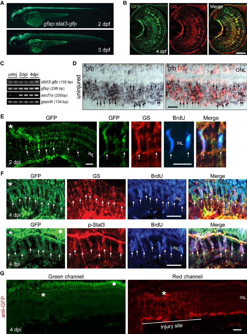
Fig. S2 Stat3-GFP expression in developing and adult gfap:stat3-gfp fish. Related to Figure 1. (A) Stat3-GFP is expressed throughout brain and spinal cord in live gfap:stat3-gfp larva at 2 and 3 dpf. (B) Immunofluorescence on retinal sections shows co-localization of Stat3-GFP with glutamine synthetase (GS)+ MG at 4 dpf. (C) Whole retina RT-PCR showing constitutive gfp mRNA expression in the uninjured and injured adult retina, while ascl1a mRNA was induced after retinal injury. (D) In situ hybridization and immunofluorescence shows expression of gfp RNA in GS+ MG in adult uninjured retina (arrows). (E) Immunofluorescence shows Stat3-GFP expression co-localizes with GS +/BrdU+ MG-derived progenitors localized to the injury site at 2 dpi in the adult retina. (F) Immunofluorescence shows co-localization of Stat3-GFP with GS+/p-Stat3+/BrdU+ MG-derived progenitors at 4 dpi in the adult retina. White dot indicates autofluorescence. Asterisks mark the injury site (needle poke) and arrows point to MG-derived progenitors. (G) Anti-GFP immunofluorescence on a retinal section prepared at 4 dpi from gfap:stat3-gfp fish using a secondary antibody coupled to a red fluor shows autofluorescence in the ONL when viewed in the green channel (left-hand panel, dot marks autofluorescence) that is not evident in the red channel (right-hand panel). Asterisk marks the injury site. Scale bar, 50 µm (B, D-G). INL, inner nuclear layer; dpf, days post fertilization.