
|
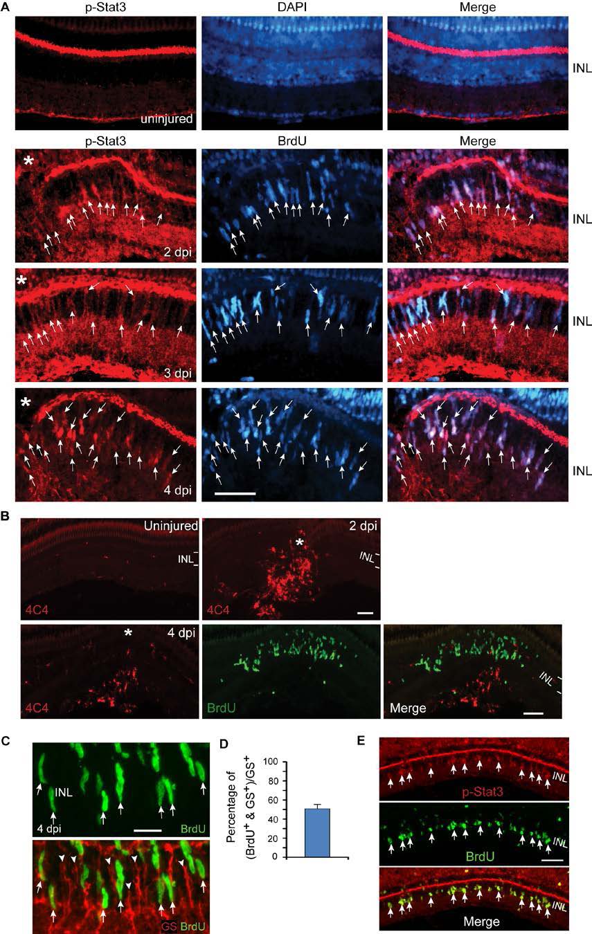
Fig. S1 Injury-dependent activation of the Jak/Stat3 signaling pathway. Related to Figure 1. (A) p-Stat3 (red) and BrdU (blue) immunofluorescence shows p-Stat3 is induced in BrdU+ MG-derived progenitors at the injury site at 2-4 dpi; n=3. Arrows point to p-Stat3+/BrdU+ double labelled cells. (B) 4C4 immunoflourescence (red) shows microglia, diffusely scattered throughout the uninjured retina, accumulate at the injury site, but do not proliferate (lack BrdU co-labeling, green). The BrdU+ cells (green) are MG-derived progenitors confined to the INL. (C, D) Immunofluorescence shows ~50% of glutamine synthetase (GS)+ MG (red) incorporate BrdU+ 4 days after a 30 min exposure to UV light; n=3. Arrows point to BrdU+/GS+ double labelled cells. Arrowheads point to BrdU-/GS+ quiescent MG. (E) Immunofluorescence shows p-Stat3 is restricted to BrdU+ MG-derived progenitors 4 days after a 30 min exposure to UV light. Asterisks in (A) point to the injury site (needle poke). Scale bar, 20 µm (C); 50 µm (A,E). INL, inner nuclear layer.