
|
Fig. 1
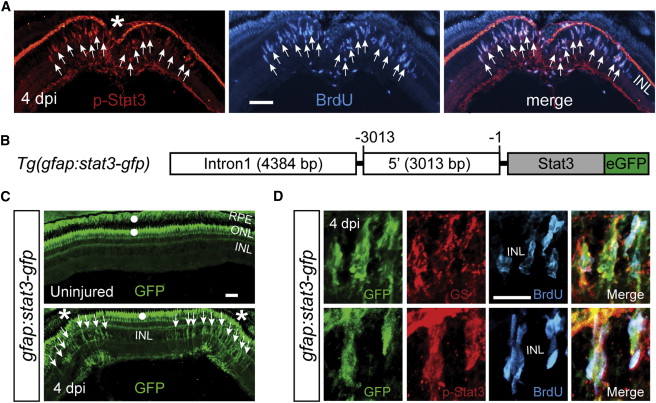
The Jak/Stat3 Signaling Pathway Is Activated following Retinal Injury
(A) Immunofluorescence on retinal sections shows activated p-Stat3 expression in BrdU+ MG-derived progenitors that are localized to the injury site at 4 dpi.
(B) A schematic of the gfap:stat3-gfp transgene construct shows the fusion gene, stat3-gfp, under control of the gfap promoter regulatory elements.
(C) In gfap:stat3-gfp transgenic fish, Stat3-GFP fusion protein expression is undetectable in MG of the uninjured eye and is restricted to MG-derived progenitors at the injury site at 4 dpi. White dots indicate autofluorescence unique to the green channel (see Figure S2G).
(D) Confocal images show colocalization of Stat3-GFP with GS+/p-Stat3+/BrdU+ MG-derived progenitors at 4 dpi.
In (A) and (C), the asterisk marks the injury site (needle poke) and arrows point to MG-derived progenitors. Scale bars, 50 µm (A and C) and 20 µm (D). INL, inner nuclear layer; ONL, outer nuclear layer; RPE, retinal pigment epithelium; dpi, days postinjury. See also Figures S1 and S2.