
|
Fig. S2
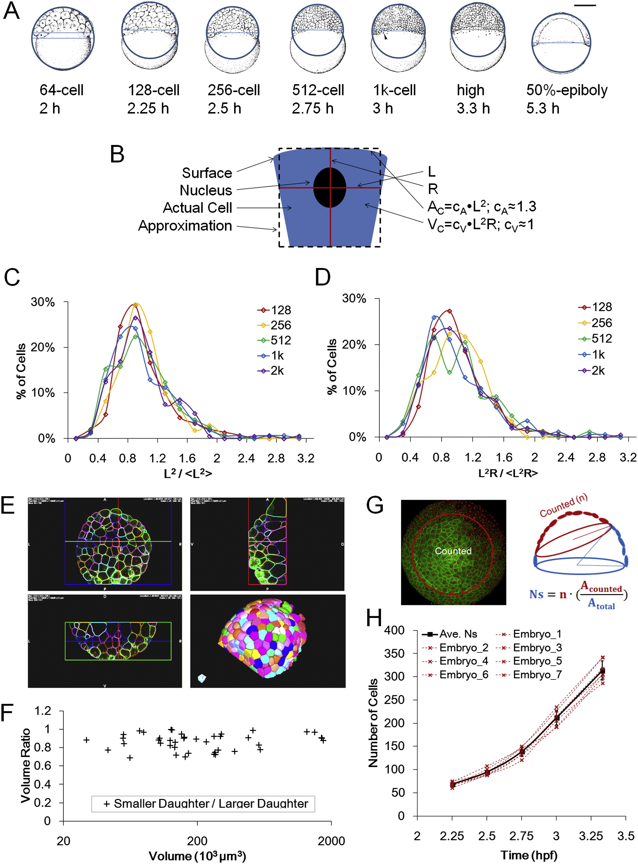
Measurements and Distributions of Surface Area, Cell Volume, and Cell Number, Related to Figure 2
(A) Schematic illustration of macroscopic surface area and volume measurement using whole embryo lateral images. Both illustrations and actual photographs (not shown) are used. The image is fit with a circle to approach the surface shape and a chord is drawn on the vegetal edge of surface cells. The surface area and volume of the resulting spherical cap provides rough whole embryo estimate values of A and VC. Embryo illustrations courtesy of Kimmel et al. (1995). See also Data S1, Text 3.
(B) Schematic of microscopic estimation of AC and VC. See also Data S1, Text 3.
(C) Distribution of normalized L2. The distribution fits a log-normal model.
(D) Distribution of normalized L2R. The distribution fits a log-normal model.
(E) Examples of full membrane segmentation to measure VC using ACME and GoFigure 2. See also Movie S2 and Extended Experimental Procedures.
(F) Ratio of sister cell volumes (smaller/larger). Data points used full membrane segmentation measurements as in (E). 7.3% ± 5.2% difference from average VC of two daughters, n = 43 pairs.
(G) Schematic illustration of surface cell counting method. Due to the spherical curvature and imaging limitations only partial surfaces were acquired, and thus an estimation of NS was made by drawing a circle on 3D perspective views of the surface. The total number equals the cell number in the circle multiplied by the ratio between surface area of the circled cap and the whole surface.
(H) Individual NS counts of 7 embryos and average. The average values were used in Figure 2 and following modeling work. “Embryo_7” is counted from a movie by Keller et al. (2010).
Reprinted from Cell, 159, Xiong, F., Ma, W., Hiscock, T.W., Mosaliganti, K.R., Tentner, A.R., Brakke, K.A., Rannou, N., Gelas, A., Souhait, L., Swinburne, I.A., Obholzer, N.D., Megason, S.G., Interplay of Cell Shape and Division Orientation Promotes Robust Morphogenesis of Developing Epithelia, 415-427, Copyright (2014) with permission from Elsevier. Full text @ Cell