
Image
|
Figure Caption
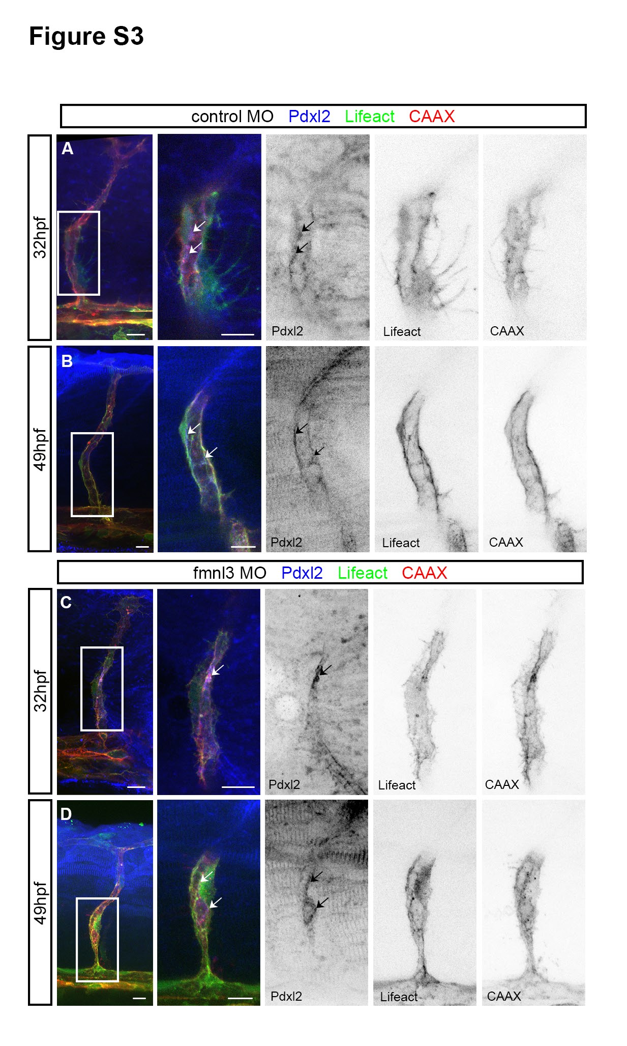
Fig. S3 Apical domains are formed in fmnl3 morphants. Related to Figure 3. (A – D) Podocalyxin-like 2 (pdxl2) staining in 32 and 49 hpf Tg(fli1ep:Lifeact- EGFP);Tg(kdr-l:ras-Cherry)s916 embryos injected with control or fmnl3 morpholinos. Arrows, apical domains. Scale bars, 10µm.
Acknowledgments
This image is the copyrighted work of the attributed author or publisher, and
ZFIN has permission only to display this image to its users.
Additional permissions should be obtained from the applicable author or publisher of the image.
Reprinted from Developmental Cell, 32, Phng, L.K., Gebala, V., Bentley, K., Philippides, A., Wacker, A., Mathivet, T., Sauteur, L., Stanchi, F., Belting, H.G., Affolter, M., Gerhardt, H., Formin-mediated actin polymerization at endothelial junctions is required for vessel lumen formation and stabilization, 123-32, Copyright (2015) with permission from Elsevier. Full text @ Dev. Cell