
|
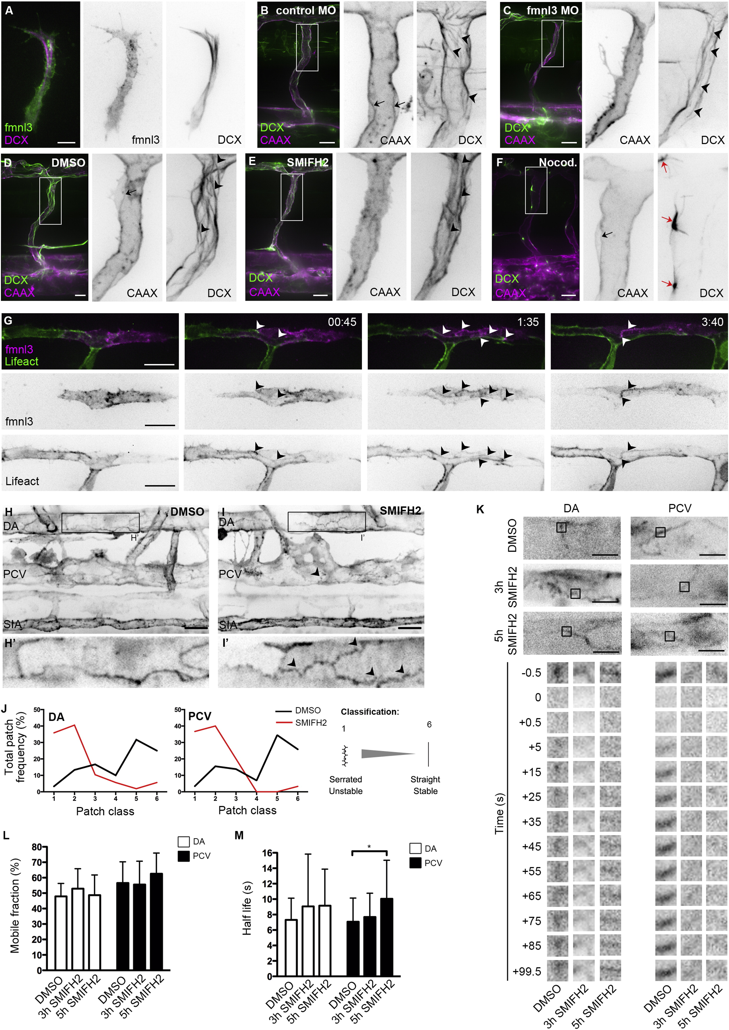
Fig. 2
Formin Activity Promotes F-Actin Polymerization at EC Junctions
(A) Coexpression of fmnl3-EGFP and DCX-mCherry in ISV at 32 hpf. Scale bar represents 10 µm.
(B and C) ISVs of Tg(fli1ep:DCX-EGFP);Tg(kdr-l:ras-Cherry)s916 embryos injected with control or fmnl3 morpholino at 49 hpf.
(D–F) Tg(fli1ep:DCX-EGFP);Tg(kdr-l:ras-Cherry)s916 embryos were treated with DMSO, 10 µM SMIFH2, or 0.5 µg/ml nocodazole at 48 hpf for 2 hr and imaged at 51–52 hpf. Arrowheads show microtubule filaments. Black arrows show apical membrane, and red arrows show microtubule organizing center. Scale bars represent 20 µm.
(G) Mosaic endothelial fmnl3-mCherry expression in Tg(fli1ep:Lifeact-EGFP) embryo from 33 hpf. Arrowheads show localization of fmnl3 with F-actin at cell junctions. Scale bar represents 20 µm.
(H–J) 3 to 4 dpf Tg(fli1ep:Lifeact-EGFP) embryos were treated with DMSO or 10 µM SMIFH2 for 4–5 hr. Arrowheads show serrated F-actin cables. DA, dorsal aorta. PCV, posterior cardinal vein. SIA, subintestinal artery. Scale bars represent 20µm. (J) Image analysis of F-actin cable profile at junctions of the DA or PCV.
(K–M) Fluorescence recovery of EGFP-Actin at cell junctions after photobleaching. Three dpf Tg(fli1ep:EGFP-Actin) embryos were treated with DMSO for 5 hr or 10 µM SMIFH for 3 or 5 hr prior to photobleaching. Scale bar represents 10 µm. Plots of EGFP-Actin mobile fraction (L) and half-life (M) are shown from different treatments. Data represent mean ± SD.
See also Figure S2.
Reprinted from Developmental Cell, 32, Phng, L.K., Gebala, V., Bentley, K., Philippides, A., Wacker, A., Mathivet, T., Sauteur, L., Stanchi, F., Belting, H.G., Affolter, M., Gerhardt, H., Formin-mediated actin polymerization at endothelial junctions is required for vessel lumen formation and stabilization, 123-32, Copyright (2015) with permission from Elsevier. Full text @ Dev. Cell