
|
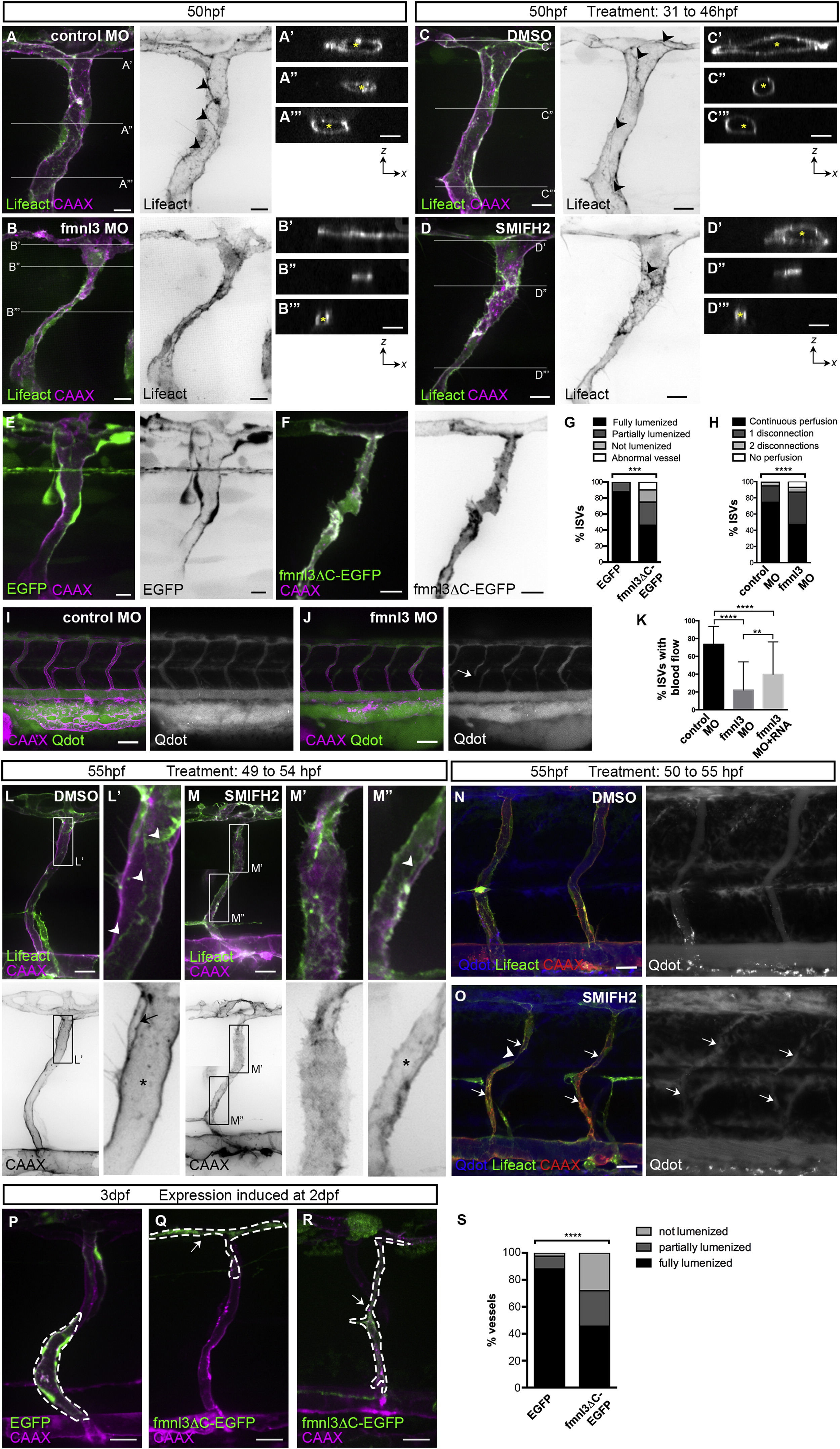
Fig. 1
Formin Activity Is Required for Vessel Lumen Formation and Maintenance
(A–D) Tg(fli1ep:Lifeact-EGFP);Tg(kdr-l:ras-Cherry)s916 embryos were injected with control or fmnl3 morpholino or treated with DMSO or 5 µM SMIFH2 from 31 to 46 hpf and examined at 50 hpf. The asterisk shows lumenized ISV, and arrowheads show junctional F-actin cables. Scale bars represent 10µm.
(E–G) Mosaic expression of EGFP or fmnl3ΔC-EGFP in ISVs of Tg(kdr-l:ras-Cherry)s916 embryos at 52 hpf. ISVs with EGFP or fmnl3ΔC-EGFP expression were phenotyped for lumen defects (G). EGFP, n = 41 ISVs, n = 16 embryos; fmnl3ΔC-EGFP, n = 52 ISVs, n = 27 embryos. Scale bars represent 10 µm.
(H–J) Tg(kdr-l:ras-Cherry)s916 embryos were injected with quantum dots (Qdot) at 54–57 hpf. The arrow shows discontinuous perfusion. Control MO, n = 134 ISVs, n = 28 embryos; fmnl3 MO, n = 186 ISVs, n = 36 embryos. Scale bars represent 50 µm.
(K) Quantification of blood flow through ISVs. Control MO, n = 79 embryos; Fmnl3 MO, n = 87 embryos; Fmnl3 MO + 100pg fmnl3 mRNA, n = 64 embryos. Data represent mean ± SD.
(L–O) Uninjected or Qdot-injected Tg(fli1ep:Lifeact-EGFP);Tg(kdr-l:ras-Cherry)s916 embryos were treated with DMSO or 10 µM SMIFH2 at 49–50 hpf and imaged 4–5 hr later. (L and M) Arrowheads show junctional F-actin cables. The arrow shows apical membrane, and the asterisk shows lumen. (O) Arrows show fragments of Qdot-filled vessels, and the arrowhead shows vessel disconnection. Scale bars represent 20 µm.
(P–S) Mosaic endothelial EGFP or fmnl3ΔC-EGFP expression (serrated lines) was induced in Tg(kdr-l:ras-Cherry)s916 embryos at 2 dpf and examined at 71–77 hpf for lumen defects. Arrows show unlumenized vessel. EGFP, n = 42 ISVs, n = 16 embryos; fmnl3ΔC-EGFP, n = 57 ISVs, n = 33 embryos. Scale bars represent 20 µm.
See also Figure S1.
Reprinted from Developmental Cell, 32, Phng, L.K., Gebala, V., Bentley, K., Philippides, A., Wacker, A., Mathivet, T., Sauteur, L., Stanchi, F., Belting, H.G., Affolter, M., Gerhardt, H., Formin-mediated actin polymerization at endothelial junctions is required for vessel lumen formation and stabilization, 123-32, Copyright (2015) with permission from Elsevier. Full text @ Dev. Cell