
|
Fig. 4
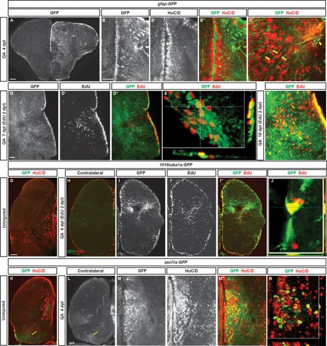
Radial glial-like progenitors respond to injury by proliferation and generation of new neurons. (A) Coronal section of the telencephalon of a QA-lesioned Tg(gfap:GFP) zebrafish shows activation of gfap manifest by GFP labeling in radial glial-like progenitors throughout the neurogenic regions in the injured hemisphere (right side of panel) in contrast to the low level of constitutive activity in the uninjured hemisphere at 4 dpl. (B–B′′) Expanded GFP expression (green) is seen in radial glial-like progenitors located in the VZ as well as hypertrophic GFP-expressing processes within the parenchyma in response to QA-induced lesion. HuC/D in red. (C) Double immunostaining for GFP (green) and HuC/D (red) in QA-lesioned brains at 4 dpl shows clusters of HuC/D+ neurons in close proximity to GFP+ processes that extend from the medial VZ to the injury (arrows indicate examples). These processes terminate in structures resembling endfeet near the lesion location (arrowhead). (D–D32) By 7 dpl, horizontal sections show that proliferative GFP+ (green) radial glial-like progenitors that incorporated EdU (red) 5 days earlier persist in the VZ, while EdU-labeled cells that appear to have migrated from the VZ to the injury site no longer express GFP (inset magnified in D′′′). (E) Orthogonal projection shows colocalization of GFP and EdU at 7 dpl. (F) At 10 dpl, costaining for EdU and GFP indicates that many of the newborn cells have migrated substantial distances toward the injury site from the lateral VZ, appearing in close proximity to or in contact with GFP+ processes (arrows indicate examples). (G–J) Tg(tuba1a:GFP) zebrafish show minimal GFP expression (green) in the uninjured telencephalon (G), increased expression mainly in the VZ of the contralesional hemisphere at 4 dpl (H), and massively increased GFP labeling in the lesioned hemisphere (I–I3) in both the VZ and parenchyma at the injury site. Many GFP+ cells were proliferating 2 days earlier (at 2 dpl) as indicated by EdU incorporation (I′′, J). (K–N) Tg(ascl1a:GFP) fish show a low level of VZ, periventricular and caudal (yellow arrows) GFP labeling (green) in the uninjured state (K) or contralateral to QA injection at 4 dpl (L). In the injured hemisphere, GFP expression increases markedly at 4 dpl in the VZ and periventricular region, and many of the cells coexpress HuC/D (M–N; red in K, M′′, and N). Scale bar = 100 µm.