
|
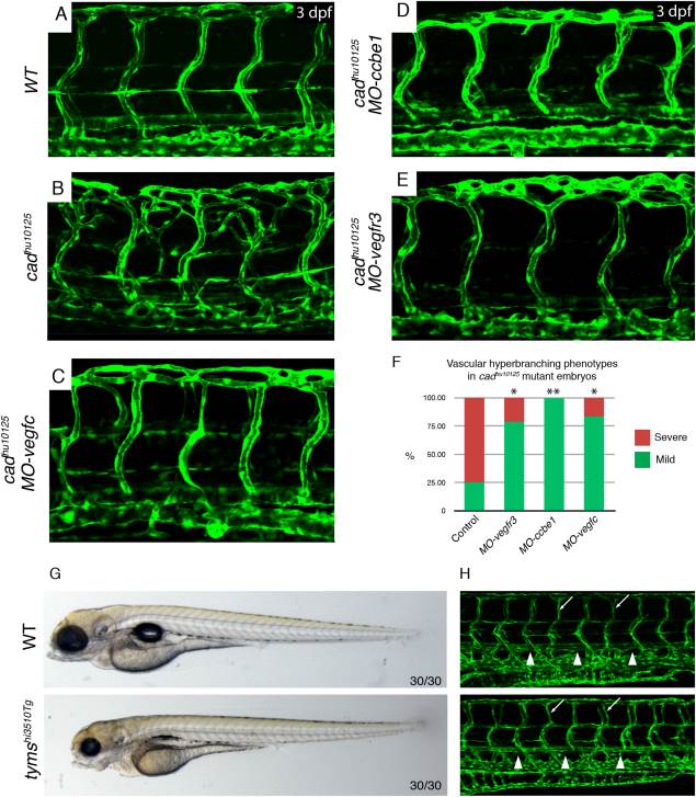
Fig. 3 cad artery hyperbranching is Vegfc/Vegfr3 dependent. A,B: The vasculature Tg(fli1a:EGFP) in wild-type sibling (A) and cadhu10125 (B) embryos at 3 dpf. C–E: The vasculature Tg(fli1a:EGFP) in cadhu10125/MO-vegfc (C), cadhu10125/MO-ccbe1 (D), and cadhu10125/MO-vegfr3 (E) embryos at 3 dpf. F: Quantification of the severity of vascular hyperbranching designated as mild or severe (where mild: 1–7 hyperbranched arteries, and severe: e 8 hyperbranched arteries along the entire embryo body axis) scored in WT (n = 16), MO-vegfc (n = 12), MO-ccbe1 (n = 10) and MO-vegfr3 (n = 14) injected cadhu10125 embryos. G,H: The thymidylate synthase mutant tymshi3510Tg does not present with arterial hyperbranching. G: Overall morphology of wild-type (n = 30/30) and tymshi3510Tg (n = 30/30) embryos at 4 dpf. H: The vasculature Tg(fli1a:EGFP) in wild-type (n = 30/30) and tymshi3510Tg (n = 30/30) embryos at 4dpf. White arrowhead: thoracic duct, white arrow: example of normal aISV.