
|
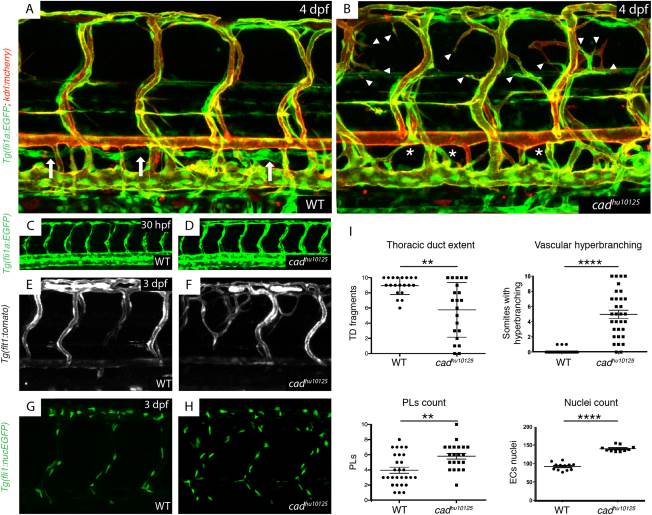
Fig. 1 hu10125 mutants present with lymphatic vascular deficiency and arterial hyperbranching. A,B: The vasculature Tg(fli1a:EGFP; flt1:tomato) in wild-type siblings (A) and hu10125 embryos (B) at 4 dpf (white arrows indicate a thoracic duct, asterisk an absence of thoracic duct, and arrowheads indicate hyperbranching). C,D: The vasculature Tg(fli1a:EGFP; flt1:tomato) in wild-type siblings (n = 53) (C) and hu10125 (n = 12) embryos (D) at 30 hpf. E,F: The arteries Tg(flt1:tomato) in wild-type siblings (C) and hu10125 (D) embryos at 3 dpf. G,H: Endothelial nuclei Tg(fli1:nucEGFP) in wild-type siblings (G) and hu10125 (H) embryos at 3 dpf. I: Quantification of: thoracic duct extent (number of thoracic duct fragments visible across 10 segments in the trunk) in WT (n = 20) and hu10125 (n = 21) (4 dpf); vascular hyperbranching (number of bilateral paired ISVs presenting with extra branches across 10 segments in the trunk) in WT (n = 32) and hu10125 (n = 32) (4 dpf); parachordal lymphangioblasts (number of PLs visible at the level of the horizontal myoseptum, across 10 segments in the trunk) in WT (n = 28) and hu10125 (n = 21) (56 hpf); and nuclei count (in ISVs, scored dorsal to the horizontal myoseptum across 10 segments) in WT (n = 12) and hu10125 (n = 10) embryos (3 dpf).