
|
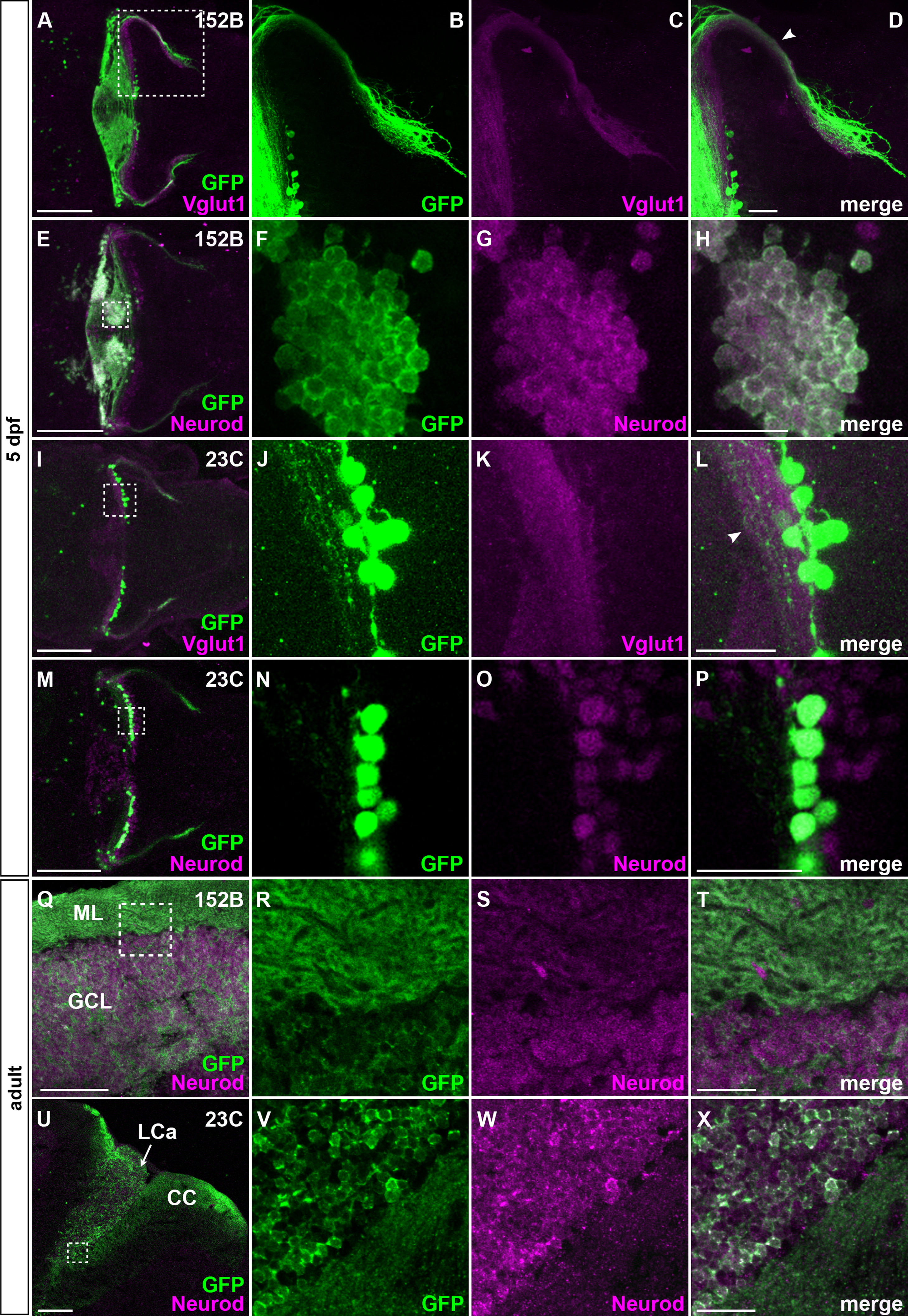
Fig. 3 Granule cell-specific GFF lines, co-staining with granule cell markers. (A–H, Q–T) gSA2AzGFF152B; UAS:GFP. (I–P, U–X) gSAIGFF23C; UAS:GFP. The 5-dpf larvae (A–P, dorsal views with anterior to the left) and sagittal sections of adult brains (Q–X, anterior to the left) were stained with anti-GFP (green) and Vglut1 or Neurod (magenta) antibodies. High magnification images of green (B, F, J, N, R, V), magenta (C, G, K, O, S, W), merged (D, H, L, P, T, X) of the box in A, E, I, M, Q, U. Projection views (A–D, E, I–L, M, Q–U) and optical sections (F–H, N–P, V–X). Note that GFP+ axons and somata were co-stained with anti-Vglut1 (arrowheads in D, L) and Neurod antibodies, respectively. There are Neurod+ and GFP- cells observed in the lobus caudalis of 5-dpf gSAIGFF23C; UAS:GFP cerebellum (Fig. 3N–P), indicating that gSAIGFF23C line marks most but not all granule cells in the lobus caudalis cerebelli. Scale bars: 100 µm in A, E, I, M; 100 µm in Q, U; 20 µm in D, H, L, P (applied to B–D, F–H, J–L, N–P, respectively); 20 µm in T, X (applied to R–T, V–X, respectively).
Reprinted from Developmental Biology, 397(1), Takeuchi, M., Matsuda, K., Yamaguchi, S., Asakawa, K., Miyasaka, N., Lal, P., Yoshihara, Y., Koga, A., Kawakami, K., Shimizu, T., Hibi, M., Establishment of Gal4 transgenic zebrafish lines for analysis of development of cerebellar neural circuitry, 1-17, Copyright (2015) with permission from Elsevier. Full text @ Dev. Biol.