
|
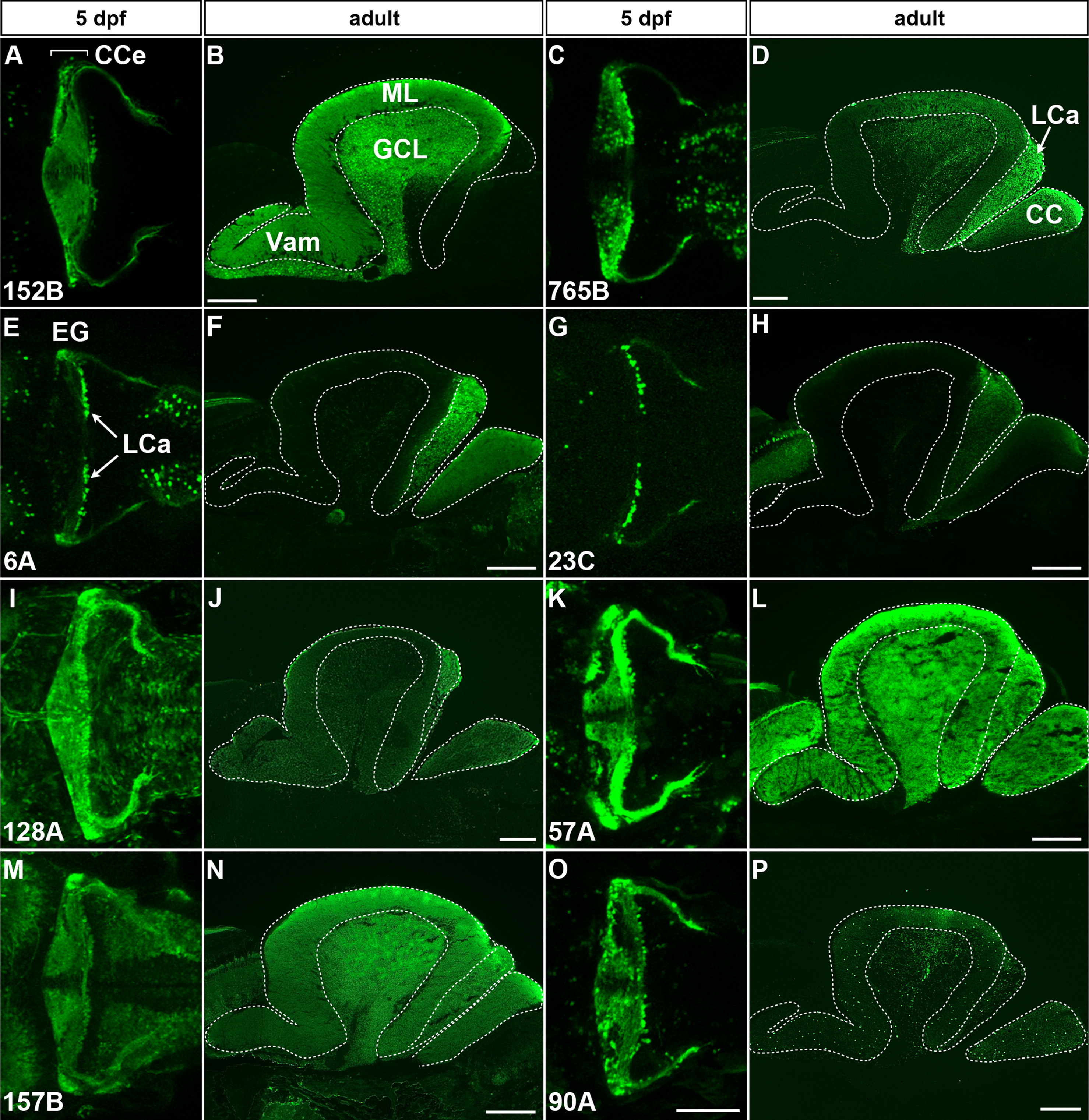
Fig. 2 Granule-specific GFF and GFP transgenic lines. (A, B) gSA2AzGFF152B; UAS:GFP. (C, D) gSAIzGFFM765B; UAS:GFP. (E, F) gSAG6A. (G, H) gSAIGFF23C; UAS:GFP. (I, J) SAGFF(LF)128A; UAS:GFP. (K, L) hspGFF57A; UAS:GFP. (M, N) SAGFF(LF)157B; UAS:GFP. (O, P) hspGFFDMC90A; UAS:GFP. To visualize GFF expression, the granule-specific GFF lines (except gSAG6A in E, F) were crossed with the UAS:GFP line. The 5-dpf larvae (A, C, E, G, I, K, M, O, dorsal projection views with anterior to the left) and sagittal sections of adult brains (B, D, F, H, J, L, N, P, optical sections with anterior to the left) were stained with anti-GFP antibody. Note that in the gSA2AzGFF152B; UAS:GFP line GFP was specifically expressed in the granule cells of the CCe, while in the gSAIzGFFM765B; UAS:GFP line GFP was expressed in some granule cells in the CCe and most granule cells in the LCa in adult. The gSAG6A and gSAIGFF23C; UAS:GFP lines expressed GFP in granule cells primarily in the LCa. In the hspGFFDMC90A; UAS:GFP line GFP was expressed in some granule cells in the GCL and LCa, and in some immature granule cells in the ML. Scale bars: 100 µm in O (applied to A, C, E, G, I, K, M, O); 200 µm in B, D, F, H, J, L, N, P.
Reprinted from Developmental Biology, 397(1), Takeuchi, M., Matsuda, K., Yamaguchi, S., Asakawa, K., Miyasaka, N., Lal, P., Yoshihara, Y., Koga, A., Kawakami, K., Shimizu, T., Hibi, M., Establishment of Gal4 transgenic zebrafish lines for analysis of development of cerebellar neural circuitry, 1-17, Copyright (2015) with permission from Elsevier. Full text @ Dev. Biol.