
|
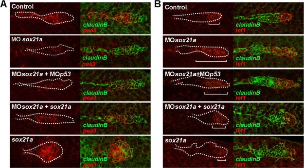
Fig. 5 Knockdown of sox21a results in a reduction of Fgf signaling and in an expansion of the Wnt signaling domain. The expression pattern in the pLL primordium of pea3 (A) or lef1 (B) is detected by fluorescent in situ hybridization in a cldnb:gfp transgenic background. Left panels represent the expression of the respective genes (red) and right panels the overlay with the GFP signal from the cldnb:gfp transgenic line (green). Different experimental conditions are shown per row, ordered top to bottom as annotated: Control (Control), sox21a morpholino background (MOsox21a), double sox21a and p53 morpholinos background (MOsox21a+MOp53), sox21a morpholino coinjected with sox21a mRNA (MOsox21a+sox21a) and injection of sox21a mRNA alone (sox21a). White dotted lines mark the borders of the pLL primordium and white brackets mark the distance from the posterior tip of the pLL primordium to the limit of expression of lef1.