
|
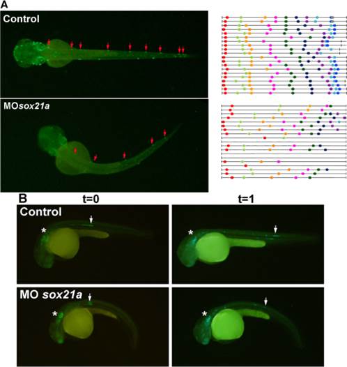
Fig. 3 Effect of sox21a knockdown in the formation of neuromasts and pLL primordium migration. A) Left panels represent dorsal images of 60hpf 3240:gfp transgenic embryos that drive expression of GFP in neuromasts (red arrows). The right panel represents the distribution of neuromasts (color dots) in different individuals (black lines). In both cases, left and right panels, above represent controls and below sox21a morphant embryos. B) Lateral images of 3240:gfp transgenic embryos acquired at two different time points, T0 (left panels; 36hpf) and T1(right panels; 42hpf). Asterisks mark the otic vesicle and arrows the pLL primordia. Controls are displayed above and sox21a morphant embryos below. The distance traveled by the pLL primordium between these two time points was measured for several embryos (n = 10 for controls and n = 10 for MOsox21a embryos).