
|
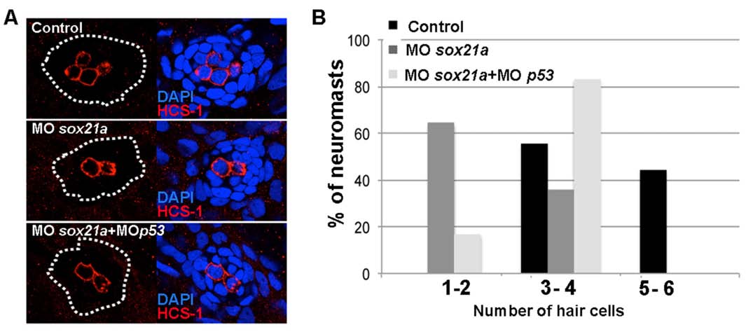
Fig. S9 Immunodetection of hair cells in differentiated neuromasts of sox21a morphant embryos. A) An anti HCS-1 antibody was used to detect hair cells (red) in differentiated neuromasts of controls (Control), sox21 morphant (MOsox21a) or sox21 and p53 double morphant embryos (MOsox21a+MOp53). Embryos were double stained with the nuclear marker DAPI (blue, right images). B) Graphs representing the percentage of analyzed neuromasts having 1 to 2, 3 to 4 or 5 to 6 differentiated hair cells, in a control (black bars; n=9), sox21a morphant (dark gray bars; n=4) or sox21a and p53 double morphant (light gray bars; n=12) backgrounds. Embryos were fixed at 72hpf.