
|
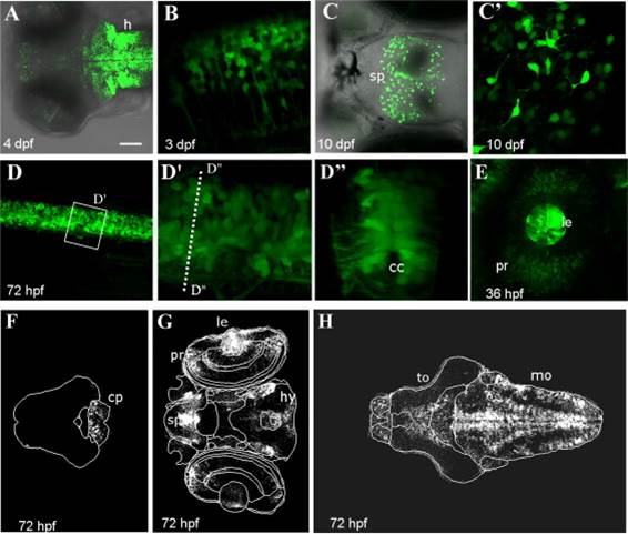
Fig. 4 Reporter expression during early larval development: central nervous system. Confocal images of brain and neural tube of Tg(12xSBE:EGFP)ia16 larvae at different stages of development. (A) Dorsal view of the brain of a 4 dpf larva (Z-stack). GFP is expressed in the hindbrain, diencephalon and telencephalon. (B) Zoomed lateral views of the hindbrain in a larva at 3 dpf (single plane). (C) Dorsal views of GFP-expressing cells in the subpallium of a 10 dpf larva (Z-stack). (C′) Zoomed dorsal view of GFP+ cells in the subpallium (Z-stack). (D) Lateral view of the neural tube in a 72 hpf larva (Z-stack). (D′–D′′), 3D-reconstruction of the neural tube of D at the level of the dashed line (D′, lateral and, D3, sagittal view). Reporter expression is mainly localized around the central canal (cc). (E) Lateral view of an eye in a 36 hpf larva (Z-stack). (F–H) Single planes of Tg(12xSBE:EGFP)ia16 brain at 72 hpf. Images have been obtained with VibeZ software. h=hindbrain, sb=subpallium, cc=central canal, pr=proliferating retina, le=lens, hy=hypothalamus, to=tectum opticum, mo=medulla oblongata. Scale bar is 100 µm in A, D; 50 µm in C; 20 µm in E; and 10 µm in B, C′, D′–D′′.
Reprinted from Developmental Biology, 396(1), Casari, A., Schiavone, M., Facchinello, N., Vettori, A., Meyer, D., Tiso, N., Moro, E., Argenton, F., A Smad3 transgenic reporter reveals TGF-beta control of zebrafish spinal cord development, 81-93, Copyright (2014) with permission from Elsevier. Full text @ Dev. Biol.