
|
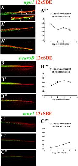
Fig. S9 Reporter expression is associated with neuronal differentiating cells. Confocal lateral view (Z-stack) of the tail of 2, 3 and 4 dpf larvae Tg(12xSBE:nls-mCherry)ia15 line with transgenic strains expressing GFP in different neuronal cells: Tg(ngn1:GFP)sb1, Tg(2.4 kb neuroD:EGFP) and Tg(mnx1:GFP)ml2. Arrowheads highlight regions of colocalization for GFP and mCherry in Tg(ngn1:GFP)sb1 and Tg(2.4 kb neuroD:EGFP): notice the coexpression at the tail tip of ngn1:GFP. The tails of each double transgenic are accompanied in the last column with a quantitative analyses of colocalization of TGFβ-mCherry expressed as Manders′ coefficient.
Reprinted from Developmental Biology, 396(1), Casari, A., Schiavone, M., Facchinello, N., Vettori, A., Meyer, D., Tiso, N., Moro, E., Argenton, F., A Smad3 transgenic reporter reveals TGF-beta control of zebrafish spinal cord development, 81-93, Copyright (2014) with permission from Elsevier. Full text @ Dev. Biol.