
|
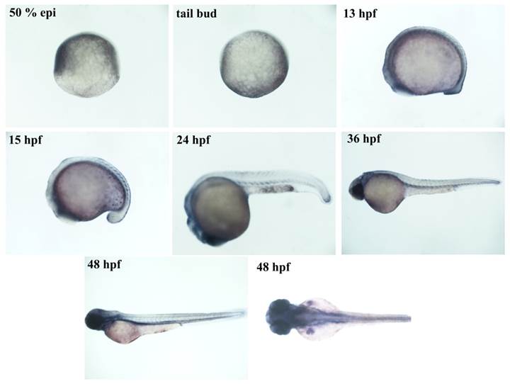
Fig. S7 phospho-Smad3 expression pattern correlates with reporter expression. Brightfield images of immunohistochemistry for phospho-Smad3 performed on wild-type embryos at the following developmental stages: 50 % epiboly, tail bud, 13, 15, 24, 36 and 48 hpf. During gastrulation the maternal derived transcription factor is active in the anterior part of the animal pole. It is absent at the end of the gastrulation and it appears again during late somitogenesis (13 hpf) in the tail, eyes and cardiac mesoderm. At 15 hpf it is extended to tail mesoderm. At 24 hpf a weak signal is visible in the cloaca and head. At 48 hpf tail mesoderm signal disappears, while fin buds become positive.
Reprinted from Developmental Biology, 396(1), Casari, A., Schiavone, M., Facchinello, N., Vettori, A., Meyer, D., Tiso, N., Moro, E., Argenton, F., A Smad3 transgenic reporter reveals TGF-beta control of zebrafish spinal cord development, 81-93, Copyright (2014) with permission from Elsevier. Full text @ Dev. Biol.