
|
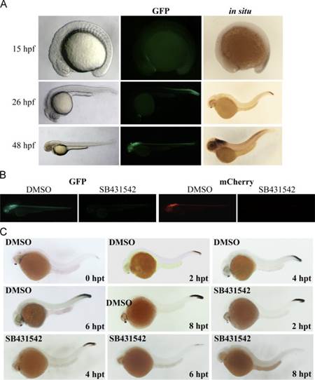
Fig. 1 D12xSBE lines are reporters of TGFβ signaling. (A) Brightfield, fluorescence and in situ hybridization lateral views of Tg(12xSBE:EGFP)ia16 embryos at 15, 26 and 48 hpf, anterior to the left. GFP expression appears in late somitogenesis in the tail and cardiac mesoderm region. At 26 hpf GFP is visible in the telencephalic region and expressed in the embryo neural tube and tail. At 48 hpf reporter expression is extended to the entire neural tube, maintaining a decreasing gradient from the tail, and in some areas of the brain. Transcription (in situ) and translation (GFP) patterns are coherent. (B) Fluorescent images of 3 dpf larvae of Tg(12xSBE:EGFP)ia16 (in green) and Tg(12xSBE:nlsmCherry)ia15, (in red) either treated with the Alk4- and Alk5-inhibitor SB431542 or with carrier (DMSO) for two days: lateral views, anterior to the left. C. RNA in situ hybridization for EGFP mRNA performed in embryos of Tg(12xSBE:EGFP)ia16 treated at 24 hpf with the Alk4- and Alk5-inhibitor, SB-431542 and fixed at different time points: 2, 4, 6, 8 h post treatment (hpt).
Reprinted from Developmental Biology, 396(1), Casari, A., Schiavone, M., Facchinello, N., Vettori, A., Meyer, D., Tiso, N., Moro, E., Argenton, F., A Smad3 transgenic reporter reveals TGF-beta control of zebrafish spinal cord development, 81-93, Copyright (2014) with permission from Elsevier. Full text @ Dev. Biol.