
|
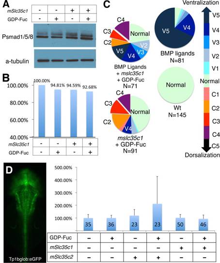
Fig. 3 BMP and Notch signaling are intact in mSlc35c1 expressing embryos. (A) Phospho-Smad1/5/8 antibody blot indicates BMP signaling activity in each treatment. (B) Quantification of Phospho-Smad1/5/8 levels, normalized to α-Tubulin protein levels. (C) Molecular interaction between BMP signaling and Slc35c1 function. (D) Dorsal view of a transgenic zebrafish larva expressing a GFP reporter of Notch activity. Graph shows the GFP expression levels determined by the fluorescence intensities of transgenic notch reporter embryos injected with the specified molecules. The data represent the average of GFP reporter intensity from individual treated embryos, with WT set as 100%.
Reprinted from Developmental Biology, 395(2), Feng, L., Jiang, H., Wu, P., Marlow, F.L., Negative feedback regulation of Wnt signaling via N-linked fucosylation in zebrafish, 268-86, Copyright (2014) with permission from Elsevier. Full text @ Dev. Biol.