
|
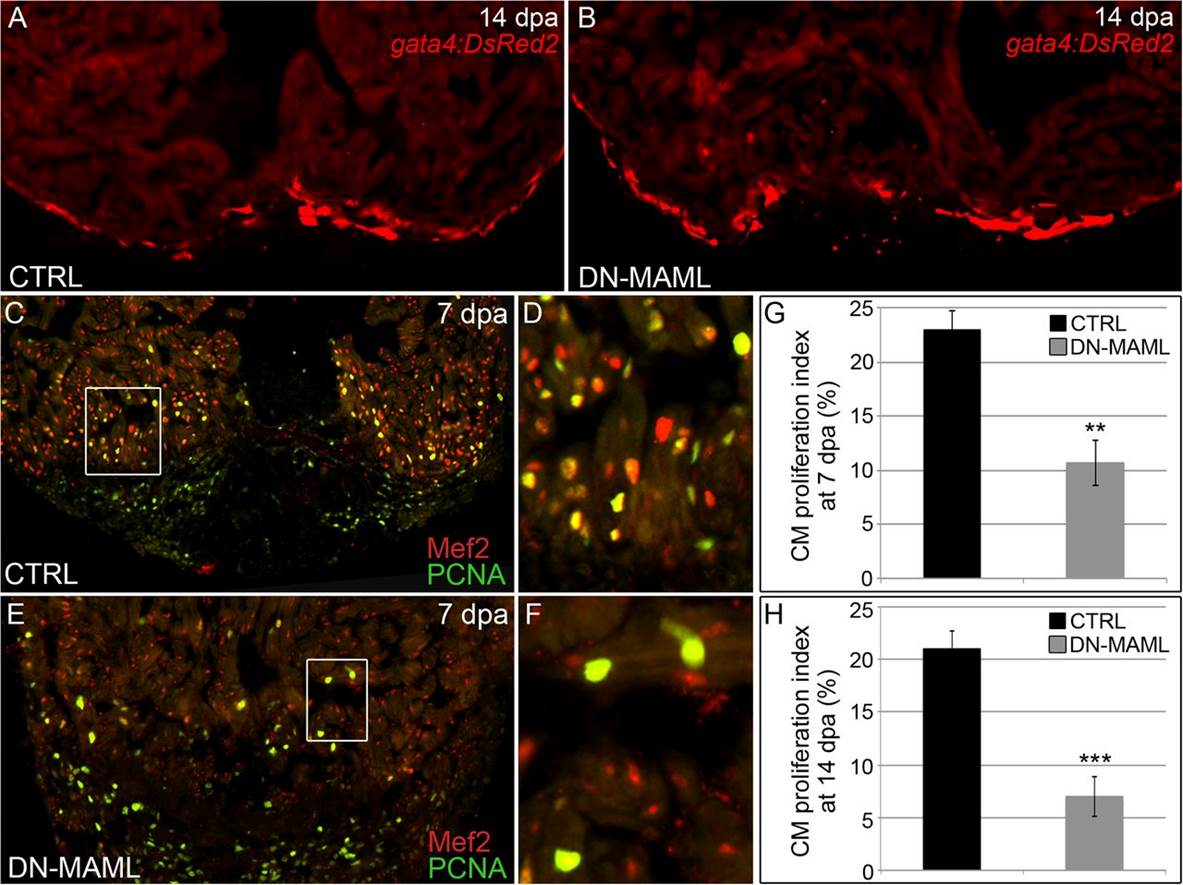
Fig. 6
Notch signaling is required for cardiomyocyte proliferation during zebrafish heart regeneration. (A and B) Representative cardiac sections from heat shocked control (CTRL) (A) and Tg(hsp70:DN-MAML) (B) animals carrying the Tg(gata4:DsRed2) transgene at 14 dpa. DsRed2 expression was visible in the compact myocardium of both control and Tg(hsp70:DN-MAML) hearts (n = 12/12 in each group). (C–F) Representative cardiac sections from heat shocked control (C and D) and Tg(hsp70:DN-MAML) (E and F) animals at 7 dpa. Sections were double immunostained to identify cardiomyocyte nuclei (Mef2+) and nuclei undergoing DNA replication (PCNA+). Boxed regions in C and E are shown at higher zoom in D and F. The percentages of myocardial nuclei undergoing DNA replication near the wound edges were quantified at 7 (G) and 14 (H) dpa and reported as mean proliferation indices ± 1 SD, **P < 0.01 and ***P < 0.001. In the control group, seven and eight hearts were analyzed at 7 and 14 dpa, respectively. In the Tg(hsp70:DN-MAML) group, three and four hearts were analyzed at 7 and 14 dpa, respectively.