
|
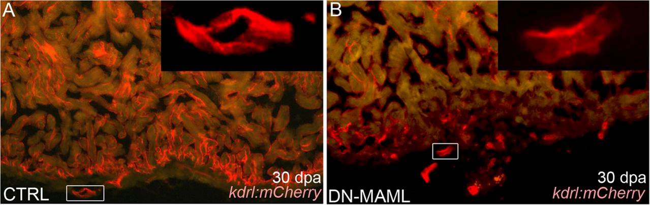
Fig. 4
Evidence for regeneration of coronary endothelium in Notch-inhibited hearts. (A and B) Representative cardiac sections from heat shocked control (CTRL) (A) and Tg(hsp70:DN-MAML) (B) animals carrying the Tg(kdrl:mCherry) endothelial reporter at 30 dpa. (Insets) Higher magnification views of boxed regions in A and B. Whereas control animals regenerated mCherry+ endocardium and coronary vessels, Tg(hsp70:DN-MAML) hearts harbored mCherry+ endothelial tubes in the nonregenerated region of the heart. The myocardium is autofluorescent in A and B. Fourteen of 14 control and 11 of 11 Tg(hsp70:DN-MAML) hearts demonstrated these phenotypes.