
|
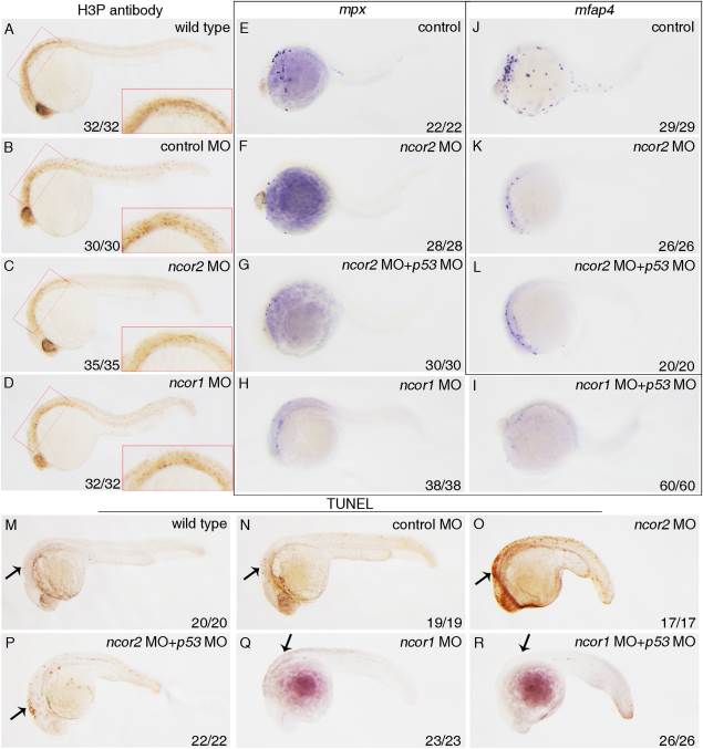
Fig. 3 The developmental defects of myeloid cells in either ncor2 or ncor1 morphants are not attributed to cell proliferation or apoptosis. A–D: Cell proliferation was detected by whole-mount in situ immunohistochemistry using phosphohistone-H3 antibody in wild-type (A), control MO (B), ncor2 MO (C), and ncor1 MO (D) embryos at 24 hpf. The inserted panels shows the head regions of embryos at higher magnification. E–L: Preventing embryonic cells from apoptosis with p53 morpholino does not rescue the defects of primitive myelopoiesis in ncor2 or ncor1 morphants. The expression of mpx (E–I) or mfap4 (J–L) was detected by whole-mount in situ hybridization when microinjected with control MO (E,J), ncor2 MO (F,K), ncor2 MO plus p53 MO (G,L), ncor1 MO (H) and ncor1 MO plus p53 MO (I), respectively. M–R: Cell apoptosis was detected by TUNEL assay in wild-type (M), control MO (N), ncor2 MO (O), ncor2 MO plus p53 MO (P), ncor1 MO (Q) and ncor1 MO plus p53 MO (R) embryos 24 hpf. Embryos were positioned anterior left and viewed laterally.