
|
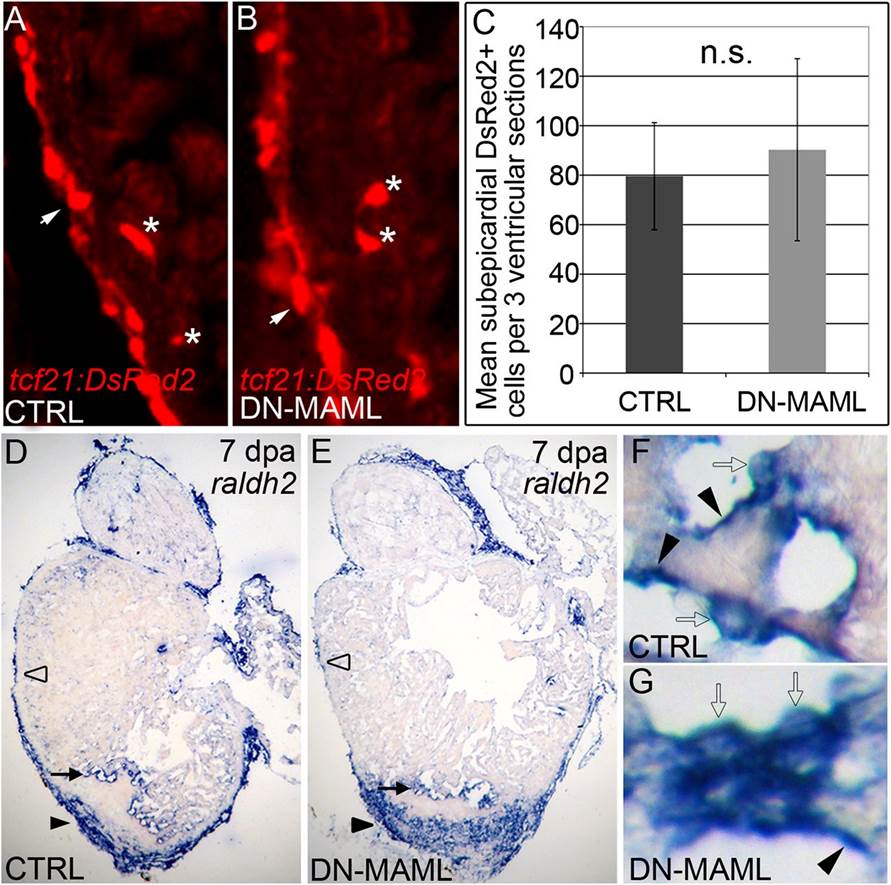
Fig. 3
Notch signaling is dispensable for injury-induced epicardial and endocardial activation. (A and B) Representative cardiac sections from 7dpa heat shocked control (CTRL) (A) and Tg(hsp70:DN-MAML) (B) animals carrying the Tg(tcf21:DsRed2) transgene. In regions away from the wound, DsRed2+ epicardial cells (arrows) and EPDCs (asterisks) were grossly normal in Notch suppressed hearts. Seven of seven hearts in each experimental group expressed DsRed2 in epicardium and EPDCs. (C) Graph representing the average numbers of subepicardial DsRed2+ cells quantified in three ventricular sections from four hearts per experimental group. n.s., not significant. (D–G) Representative cardiac sections from heat shocked control (D) and Tg(hsp70:DN-MAML) (E) animals at 7 dpa stained for raldh2 transcripts by in situ hybridization. In both experimental groups, prominent raldh2 expression was observed in epicardial cells covering the wound (black arrowheads in D and E), away from the wound (open arrowheads in D and E), and in endocardial cells near the wound edge (black arrows in D and E). Higher magnifications of endocardial raldh2 expression are shown in F and G. Endocardial cells near the wound displayed both flattened (black arrowheads in F and G) and rounded morphologies (open arrows in F and G). Four of four hearts in each experimental group expressed endocardial and epicardial raldh2 transcripts.