
|
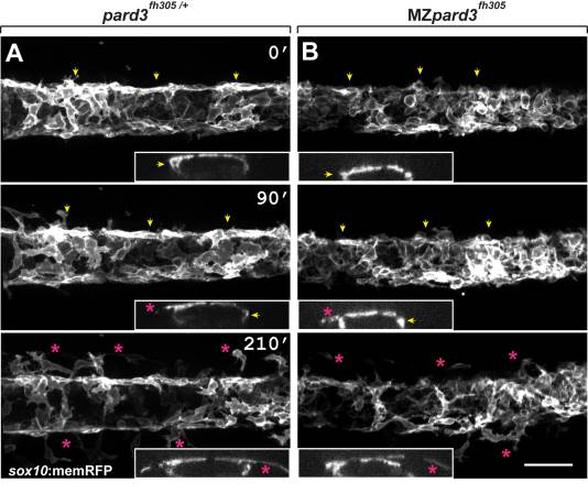
Fig. 2 Schwann cells exit the dorsal neural tube on schedule in pard3 mutants. Panels show representative frames from time-lapse movies of trunk neural crest cells between 18–24 hpf. Views are from dorsal with anterior to the left. Insets display z-plane orthogonal views of the migrating neural crest for each frame. Numbers denote time elapsed, in minutes, from start of imaging. Embryos are siblings and imaged sequentially in the same chamber. A: Initiation of neural crest migration in a control pard3fh305/+ embryo. Neural crest cells are marked by sox10:memRFP expression. Arrows mark migration on the medial pathway between neural tube and somites and asterisks mark migration on the lateral pathway, across the dorsolateral surface of somites. B: Migration of neural crest in a MZpard3fh305 mutant embryo. The onset and pattern of migration is similar to the control. Scale bar = 50 µM.