
|
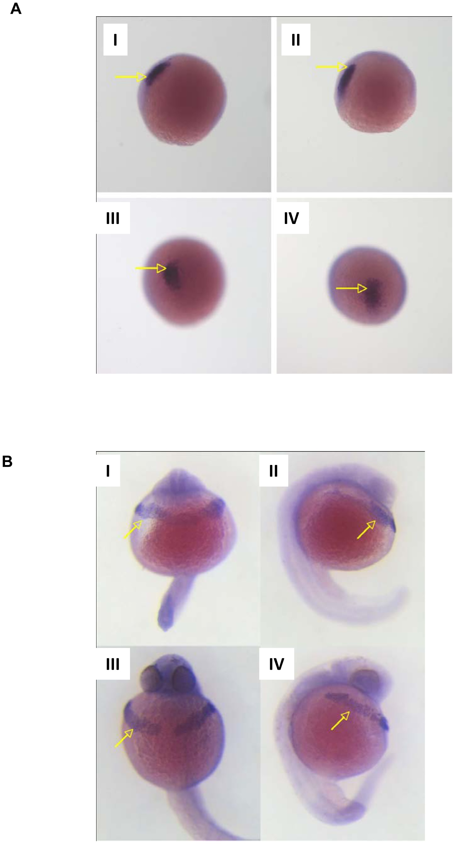
Fig. 6
Determination of mesodermal markers by ISH.
Morphant embryos were injected with 10 pg MO4. A. Location of pre-chordal plate marker gsc in LWT embryos at 80% epiboly. I and II give lateral view, III and IV looking down on gsc positive staining. II and IV were injected with MO4, I and III are mis-match injected siblings. Arrows indicate gsc specific staining. B. Hatching gland marker cat L in 24 hour post fertilisation LWT embryos. I and II are morphant embryos, ventral and lateral views respectively. III and IV are mismatch injected LWT embryos, ventral and lateral views respectively. Arrows indicate hatching gland specific cat L staining.