
|
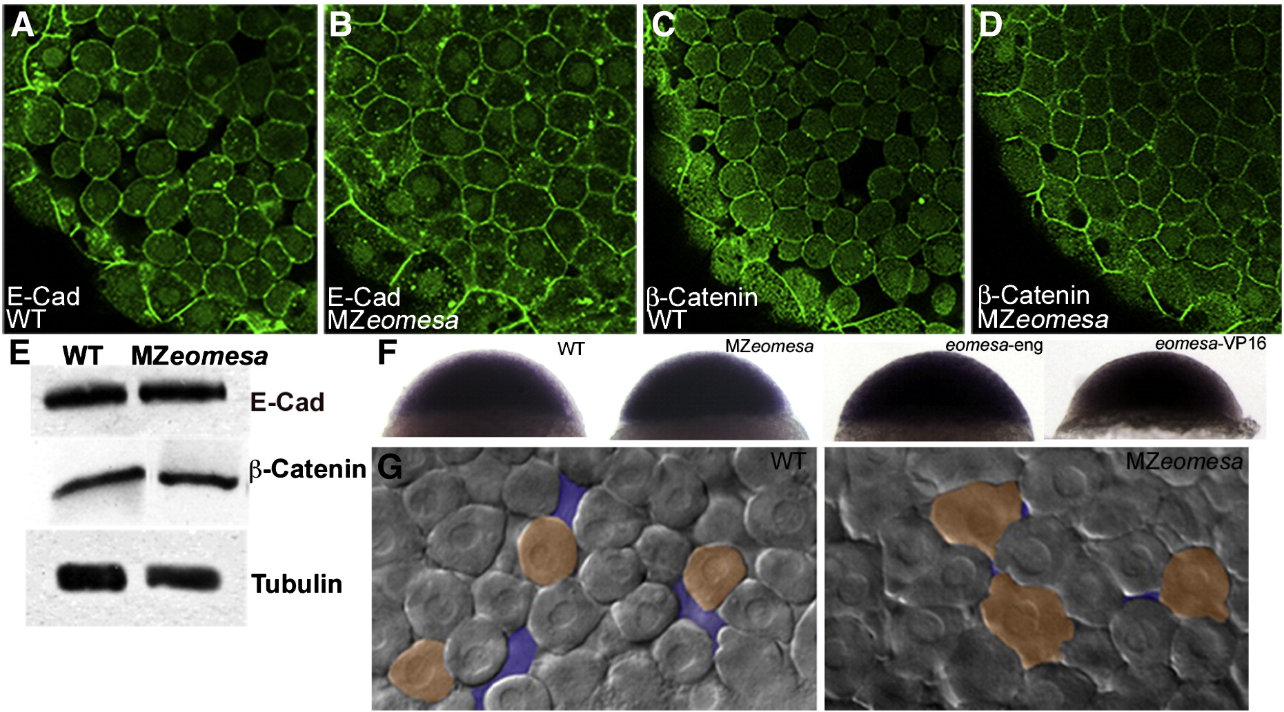
Fig. 7 Cell morphology is altered in MZeomesa embryos. (A-D) Confocal images of embryos at sphere stage stained for Cdh1 (A,B) and β-Catenin (C,D). (E) Western blot of wild type (lane 1) and MZeomesa mutant embryo (lane 2) sphere stage extracts. Levels of Cdh1 and β-Catenin are not obviously altered in mutant embryos. (F) Normal cdh1 expression in sphere stage wild type, MZeomesa, eomesa-eng and eomesa-VP16 injected wild type embryos, as indicated. (G) Live DIC images of wild type and MZeomesa embryos at sphere stage. Mutant cells are more tightly packed and exhibit more blebs then wild type cells. Orange highlights selected cell morphologies, purple highlights intercellular space.
Reprinted from Developmental Biology, 362(1), Du, S., Draper, B.W., Mione, M., Moens, C.B., and Bruce, A.E., Differential regulation of epiboly initiation and progression by zebrafish Eomesodermin A, 11-23, Copyright (2012) with permission from Elsevier. Full text @ Dev. Biol.